Melike Lakadamyali, Ph.D.
University of Pennsylvania
The Perelman School of Medicine
Department of Physiology
764 Clinical Research Building
415 Curie Blvd
Philadelphia, PA 19104
Office: 215-746-5150
Fax: 215-746-5150
Lab: 215-746-5150

- Developed versatile methods for quantitative, multiplexed imaging of molecular complexes using super-resolution microscopy:Proteins in cells assemble into nanoscale complexes in order to carry out a specific function. The spatial organization and stoichiometry of proteins within these nanoscopic functional units is highly important for maintaining a cell’s healthy physiology. Changes in nanoscale organization of protein complexes, for example a change from monomeric to oligomeric stoichiometry, often triggers disease states. However, visualizing many proteins simultaneously and quantifying protein copy number with high spatial resolution is highly challenging. Super-resolution methods hold promise for overcoming this hurdle, however, the complex photophysics of fluorophores limits both multi-color imaging capabilities and the ability to extract quantitative information. Lakadamyali lab has been developing new methods to overcome these challenges. To address the limitations in simultaneous, high-throughput, multi-color super-resolution imaging, we recently combined DNA-Paint with excitation multiplexing to demonstrate the initial proof of concept for fm-DNA-Paint. Further, to address the challenges with quantification of protein copy number at the nanoscale level, we built calibration standards based on DNA origami. Overall, these methods begin to overcome some of the main challenges associated to super-resolution microscopy making it a multiplexed and quantitative tool.
- “Quantifying protein copy number in super-resolution using an imaging invariant calibration”,F.C. Zanacchi, C. Manzo, R. Magrassi, N.D. Derr,M. Lakadamyali, Biophysical Journal, 116, 2195-2203 (2019)
- “Excitation-multiplexed multicolor super-resolution imaging with fm-STORM and fm-DNA-Paint” P.A. Gómez-García, E.T. Garbacik, M.F. Garcia-Parajo,M. Lakadamyali,PNAS, 115, 12991-12996 (2018)
- “DNA Origami: Versatile super-resolution calibration standard for quantifying protein copy number” F. Cella Zanacchi, C. Manzo, A. Sandoval Alvarez, N, Derr,M. Lakadamyali,Nature Methods, doi:10.1038/nmeth.4342 (2017)
- “Single molecule evaluation of fluorescent protein photoactivation efficiency using anin vivonanotemplate”, N. Durisic, L. L. Cuervo, A. S. Álvarez, J.Borbely, M. Lakadamyali, Nature Methods, 11, 156-162 (2014)
- Determined a novel nanoscale organization of chromatin:An important goal of the Lakadamyali lab is to reconcile the epigenomic and microscopic views of chromatin organization and determine how chromatin structure regulates gene function. Nuclear organization of the chromatin fiber spans many length-scales and while the nanoscale level organization plays a key role in regulating gene expression, this organization is impossible to visualize using conventional microscopy methods. Lakadamyali lab has been using and further developing quantitative super-resolution microscopy methods to overcome this limitation. We visualized and estimated the number of nucleosomes along the chromatin fiber of different cells at nanoscale resolution. We discovered that nucleosomes are assembled in heterogeneous groups of varying sizes, which we termed “clutches”. Remarkably, the median number of nucleosomes and their packing density inside clutches highly correlated with cellular state, such that clutch size correlates with gene expression and pluripotency grade of iPSCs. Additionally, nanoscale chromatin organization is aberrantly remodeled in disease states.
- “Aberrant chromatin reorganization in cells from diseased fibrous connective tissue in response to altered chemomechanical cues”. S. Heo, S. Thakur, X. Chen, C. Loebel, B. Xia, R. McBeath, J.A. Burdick, V.B. Shenoy, R.L. Mauck, M. Lakadamyali, Nature Biomedical Engineering, in press (2022)
b. “Two-Parameter mobility assessments discriminate diverse regulatory factor behaviors in chromatin”, J. Lerner, P.A. Gomez-Garcia, R. McCarthy, Z. Liu, M. Lakadamyali, K. S. Zaret, Molecular Cell, doi: https://doi.org/10.1016/j.molcel.2020.05.036, (2020)
- “Super-resolution microscopy reveals how histone tail acetylation affects DNA compaction within nucleosomes in vivo”, J. Otterstrom, A.C. Garcia, C. Vicario, P.A. Gomez-Garcia, M.P. Cosma, M. Lakadamyali, Nucleic Acids Research, doi: 10.1093/nar/gkz593 (2019)
- “Chromatin fibers are formed by heterogeneous groups of nucleosomes in vivo”, M. A. Ricci, C. Manzo, M. Garcia-Parajo, M. Lakadamyali†, M. P. Cosma†, (†co-senior authors), Cell, 160, 1145-1158 (2015)
3. Developed cutting-edge methods for studying intracellular trafficking: One of the overarching goals of Lakadamyali lab is to determine how molecular motors coordinate to transport vesicles and organelles in the complex cellular environment. To achieve this goal, we developed an “all-optical correlative imaging method” that combines single particle tracking in living cells with super-resolution microscopy of the same cell after fixation. Super-resolution microscopy can reveal the details of cellular architecture with exquisite spatial resolution; however, the current methods cannot capture fast dynamic processes (millisecond time scale) due to limited temporal resolution. With the correlative approach, we overcame this limitation and for the first time related cargo transport dynamics to the organization of the microtubule cytoskeleton in the cellular context. By mapping cargo trajectories to individual microtubules, we studied how the local cytoskeletal architecture regulates vesicle trafficking. We found that the cytoskeleton acts as a selective filter to vesicles that are comparable in size to or larger than the mesh size of the microtubule network leading to their pausing. We further showed that lysosomal and autophagosomal compartments are selectively enriched on specific microtubule tracks defined by post-translational modifications in order to enhance their fusion and regulate autophagy. Overall, we have developed powerful methods that reveal how the 3D local cytoskeletal architecture and the microtubule post-translational modifications combine to regulate, opening a new window into studying vesicle-cytoskeleton interactions in the physiological as well as disease contexts. We have recently expanded these studies to visualizing the distribution of microtubule associated proteins like tau under physiological and pathological conditions.
- “Tau forms oligomeric complexes on microtubules that are distinct from pathological oligomers”, M.T. Gyparaki, A. Arab, E.M. Sorokina, A.N. Santiago-Ruiz, C.H. Bohrer, J. Xiao, M. Lakadamyali, PNAS, 118 (19) e2021461118 (2021)
- “Detyrosinated microtubules spatially constrain lysosomes facilitating lysosome-autophagosome fusion” N. Mohan, I. V. Verdeny, A. S. Alvarez, E. Sorokina, M. Lakadamyali, Journal of Cell Biology, doi: 10.1083/jcb.201807124, (2018)
- “3D Motion of Vesicles Along Microtubules Helps Them to Circumvent Obstacles in Cells”, I. V. Vilanova, F. Wehnekamp, N. Mohan, A. S. Alvarez, J. S. Borbely, J. Otterstrom, D. Lamb, M. Lakadamyali, Journal of Cell Science, doi: 10.1242/jcs.201178, (2017)
- “Correlative live-cell and superresolution microscopy reveals cargo transport dynamics at microtubule intersections”, Š. Bálint, I. V. Verdeny, A. S. Álvarez, M. Lakadamyali, PNAS, 110, 3375-3380 (2013) (cover page illustration)
Research Interest
The overarching goal of the Lakadamyali lab is to understand the molecular mechanisms that regulate sub-cellular organization and the significance of this organization on cell function. Cells are highly compartmentalized: the sub-cellular positioning of organelles, nucleic acids and proteins are spatially and temporally coordinated to ensure that biochemical reactions take place at the right place and time. Dr. Lakadamyali’s program has three major focus areas that seek to advance our understanding of sub-cellular organization. First, she develops advanced microscopy methods that provide the technological advances necessary to visualize the spatial organization of cellular machinery with near molecular spatial resolution, including quantitative tools for measuring protein stoichiometry and multiplexed, high-throughput imaging methods. Second, she seeks to determine how the microtubule cytoskeleton and motors regulate transport and positioning of organelles within the cytoplasm and the functional consequences of disrupting proper organelle organization. As part of this work, her lab has recently expanded their focus to studying microtubule associated protein tau and its aggregation in neurological diseases. Third, she seeks to understand how the spatial organization of chromatin within the nucleus regulates gene activity. These three areas integrate synergistically to move forward our understanding of how sub-cellular organization emerges and impacts cell physiology and pathology.
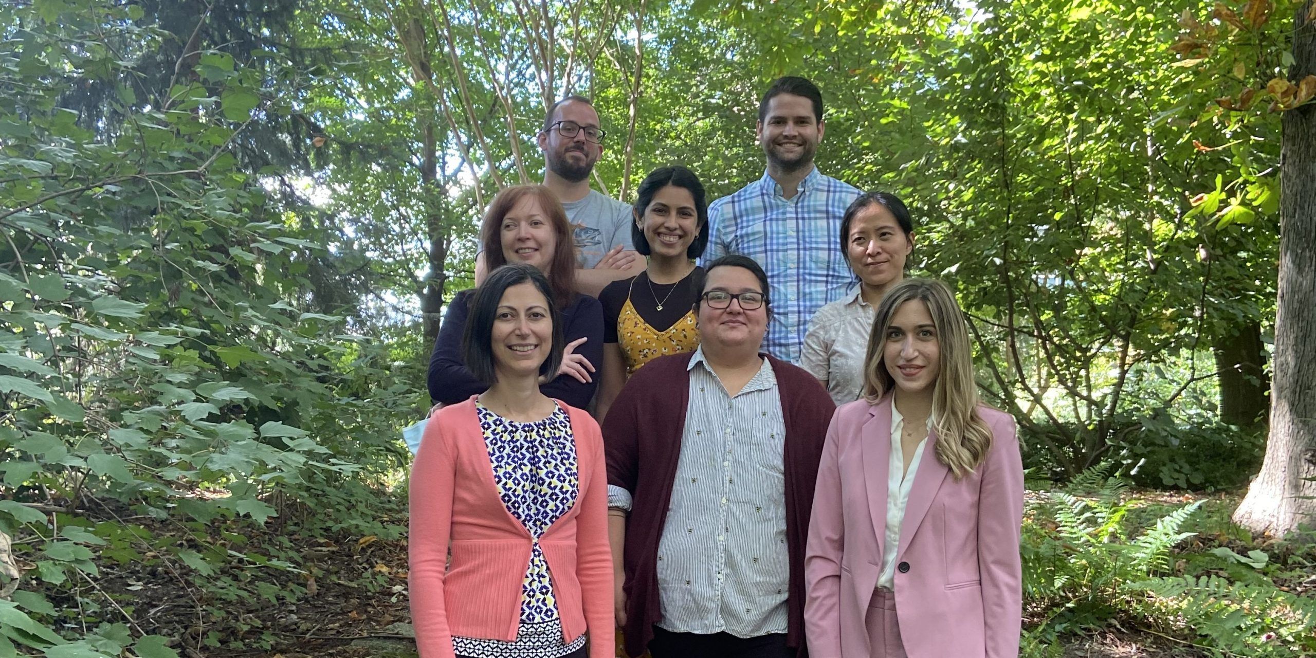
Lab Members
| First Name | Last Name | Title | |
|---|---|---|---|
| Melike | Lakadamyali | PI | melikel@pennmedicine.upenn.edu |
| Jose Angel | Martinez Sarmiento | Visiting Student, PhD Candidate | jangelmtzs@outlook.com |
| Hannah | Kim | PhD candidate | Hannah.Kim3@pennmedicine.upenn.edu |
| Marcus | Woodworth | Postdoc | marcus.woodworth@pennmedicine.upenn.edu |


