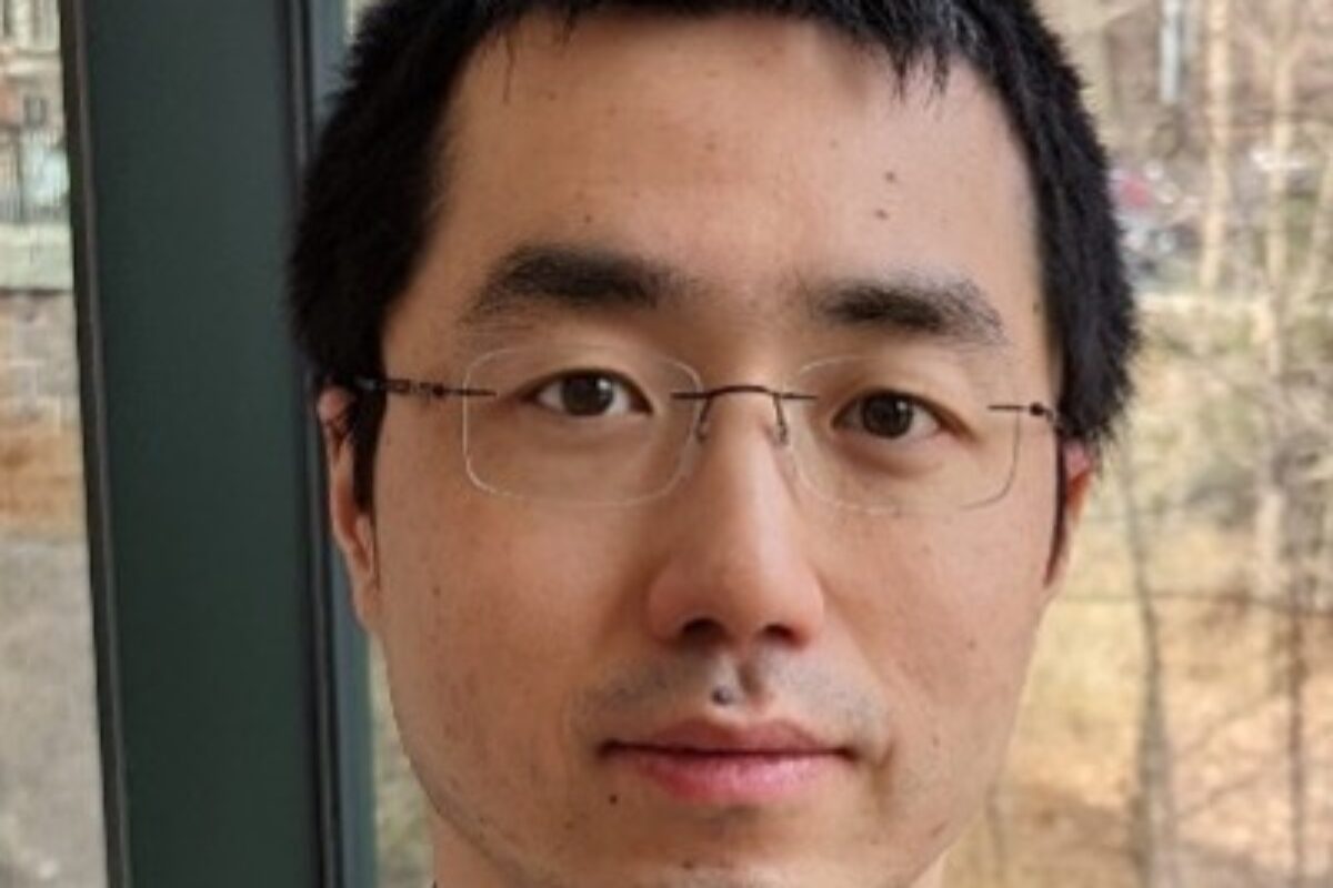
Yanxiang Deng, Ph.D.

  1. Spatially resolved profiling of histone modifications

To investigate the mechanisms underlying spatial organization of different cell types and functions in the tissue context, it is highly desired to examine not only gene expression but also epigenetic underpinnings in a spatially resolved manner to uncover the causative relationship determining what drives tissue organization and function. Despite recent advances in spatial transcriptomics to map gene expression, it has not been possible to determine the underlying epigenetic mechanisms controlling gene expression and tissue development with high spatial resolution. We developed a first-of-its-kind technology called spatial-CUT&Tag for genome-wide profiling of histone modifications pixel by pixel on a frozen tissue section without dissociation. This method resolved spatially distinct and cell-type-specific chromatin modifications in mouse embryonic organogenesis and postnatal brain development. Single-cell epigenomic profiles were derived from the tissue pixels containing single nuclei. Spatial-CUT&Tag adds a new dimension to spatial biology by enabling the mapping of epigenetic regulations broadly implicated in development and disease. In addition, epigenetic drugs are emerging now, so potentially we can develop drugs to target those epigenetic mechanisms. Having the tools to understand the epigenetic origin of different disease states could open up a whole new avenue of therapeutics.

  • Deng, Y., Bartosovic, M., Kukanja, P., Zhang, D., Liu, Y., Su, G., Enninful, A., Bai, Z., Castelo-Branco, G. and Fan, R. Spatial-CUT&Tag: spatially resolved chromatin modification profiling at the cellular level, Science, 375: 681-686 (2022).
  1. Spatially resolved profiling of chromatin accessibility

An ambitious global initiative has been undertaken to map cell types across all human organs. Single-cell sequencing has been critical to this effort, but it is hard to map the location of cell types to the original tissue environment. We developed spatial-ATAC-seq, which for the first time allows for directly observing cell types in a tissue as defined by global epigenetic state. Spatial-ATAC-seq allows us to identify which regions of the chromatin are accessible genome-wide in cells at specific locations in a tissue. This chromatin accessibility is required for genes to be activated, which then provides unique insights on the molecular status of any given cell. Combining the ability to analyze chromatin accessibility with the spatial location of cells is a breakthrough that can improve our understanding of cell identity, cell state and the underlying mechanisms that determine the expression of genes in the development of different tissues or diseases. Profiling mouse embryos using spatial-ATAC-seq delineated tissue-region-specific epigenetic landscapes and identified gene regulators involved in the development of the central nervous system. Mapping the accessible genome in the mouse and human brain revealed the intricate arealization of brain regions. Applying spatial-ATAC-seq to tonsil tissue resolved the spatially distinct organization of immune cell types and states in lymphoid follicles and extrafollicular zones. This technology progresses spatial biology by enabling spatially resolved chromatin accessibility profiling to improve our understanding of cell identity, cell state and cell fate decision in relation to epigenetic underpinnings in development and disease.

  • Deng, Y., Bartosovic, M., Ma, S., Zhang, D., Kukanja, P., Xiao, Y., Su, G., Liu, Y., Qin, X., Rosoklija, B.R, Dwork, A., Mann, J.J., Xu, M.L., Halene, S., Craft, J.E., Leong, W.K., Boldrini, M., Castelo-Branco, G. and Fan, R. Spatial profiling of chromatin accessibility in mouse and human tissues, Nature, 609: 375–383 (2022).
  1. Spatially resolved transcriptomics and proteomics

In multicellular systems, cells do not function in isolation but are strongly influenced by spatial location and surroundings. Spatial gene expression heterogeneity plays an essential role in a range of biological, physiological, and pathological processes. We developed a novel microfluidic platform (DBiT-seq) to deliver molecular barcodes to formaldehyde or FFPE fixed tissue sections in a spatially confined manner, enabling simultaneous barcoding of mRNAs and proteins, and construction of a high-spatial-resolution multi-omics atlas by NGS sequencing. The unique microfluidic in-tissue barcoding technique has enabled high-spatial-resolution mapping of whole transcriptome and tens of proteins at cellular level.

  • Liu, Y#, Yang, M#, Deng, Y#, Su, G, Enninful, A, Guo, C, Tebaldi, T, Zhang, D, Kim, D, Bai, Z, Norris, E, Pan, A, Li, J, Xiao, Y, Halene, S and Fan, R. “High-Spatial-Resolution Multi-Omics Sequencing via Deterministic Barcoding in Tissue”, Cell, 183: 1–17 (2020).

Research Interest

The Deng lab is developing novel technologies for spatial omics to solve challenging biological problems, including cancer and neurodegenerative diseases.

Kahlilia Morris-Blanco, Ph.D.

Research Interest

Dr. Morris-Blanco’s laboratory investigates epigenetic mechanisms involved in stroke pathophysiology by examining the interplay between spatial and temporal epigenetic dynamics, transcriptional regulation, and mitochondrial function in the post-stroke brain. Using both in vitro and in vivo experimental stroke models, they employ gene-specific and genome wide assessments of epigenomic organization, single-cell omics, metabolomics, and functional assessments of mitochondria and neuroprotection. Dr. Morris-Blanco is especially interested in using these mechanistic studies to develop novel treatment strategies, with the goal of translating epigenetic therapies to the clinic.

Aman Husbands, Ph.D.

Research Interest

Despite coordinating incredible morphological complexity, developmental patterning is remarkably robust. We are interested in uncovering the properties that allow complex biological processes, like development, to occur so reproducibly. One attractive system to study these ideas is the production of flat leaf architecture. The leaves of many species emerge from the stem cell niche as radially symmetric bumps, then develop into long and wide, but very shallow, structures. Leaves have solved this difficult biological problem by using the boundary between their dorsal (adaxial or top) and ventral (abaxial or bottom) sides as a guide to orient their growth. Ensuring the dorsoventral axis is rigorously specified and maintained is thus key to the robust nature of flat leaf production. We exploit the complex, gene regulatory network underlying dorsoventral patterning to assess the determinants – and their interactions – that lead to robust developmental outcomes in multicellular organisms.

A parallel but overlapping project involves the CLASS III HOMEODOMAIN LEUCINE ZIPPER (HD-ZIPIII) proteins. This ancient family of transcription factors arose at least 700 million years ago, and was repeatedly co-opted to drive several evolutionarily-important innovations, including flat leaf production, stem cell maintenance, and vascular patterning. In addition to DNA-binding and dimerization domains, HD-ZIPIII proteins contain a StAR-related transfer (START) domain, raising the intriguing possibility that HD-ZIPIII activity may be under direct control of a lipophilic ligand. Determining how HD-ZIPIII proteins are able to function in such different developmental contexts, and identifying their putative ligands, are central goals of the lab. Given their broad and deep conservation throughout the plant kingdom, we are also considering these ideas through the lens of evolution.

Mustafa A. Mir, Ph.D.

1. Label-free blood screening instruments (Masters work). Whole blood analysis is a critical and familiar part of clinical workflows. Widely used clinical blood cytometers are generally expensive and bulky, require proprietary reagents, and provide limited quantitative information on cell morphology. When these automated cytometers do find abnormalities, manual examination of a stained blood smear using light microscopy is required for further diagnosis. This process is costly, slow, and not quantitative. In my early graduate career, working Prof. Gabriel Popescu, I set out to address these issues by utilizing Quantitative Phase Imaging (QPI). In QPI the phase shift (optical delay) of light passing through a sample is measured with sub-nanometer accuracy using interferometry providing high resolution topographic and tomographic data (Mir et al, Prog. Optics, 2012). I started by building a new QPI instrument utilizing commercial CD-ROM technology (Mir et al., Opt. Express., 2009) with the goal of developing a low-cost platform for an automated point-of-care blood screening tool. A comparison of results on patient samples with those from a clinical cytometer shows excellent agreement (Mir et al., J. Biomed. Opt., 2010 & Biomed. Opt. Exp., 2011) for clinical parameters on red blood cells. Additionally, my approach provides quantitative single cell morphological data, uses 2 orders of magnitude lower input sample, requires no reagents other than whole blood, and significantly lowers instrument cost. Collectively, these studies established that QPI based technologies are more sensitive than currently used clinical whole blood analyzers for common parameters used to characterize erythrocytes and also provide additional information that could lead to earlier diagnosis of certain diseases. 

a) M. Mir, B. Bhaduri, R. Wang, R. Zhu, G. Popescu, Chapter 3 – Quantitative Phase Imaging, Progress in Optics, Ed: W. Emil, Elsevier, Volume 57, pp. 133-217 (2012) 

b) M. Mir, Z. Wang, K. Tangella and G. Popescu, Diffraction Phase Cytometry: Blood on a CD-ROM, Optics Express, 17 (4), 2579-2585 (2009) 

c) M. Mir, H. Ding, Z. Wang, J. Reedy, K. Tangella and G. Popescu, Blood Screening using Diffraction Phase Cytometry, Journal of Biomedical Optics, 15 (2), 027016 (2010) 

d) M. Mir, K. Tangella and G. Popescu, Blood Testing at the Single Cell Level using Quantitative Phase and Amplitude Microscopy, Biomedical Optics Express, 2 (12), 3259-3266. (2011) 

2. Non-perturbative, quantitative, and label-free live imaging spanning broad spatio-temporal scales (PhD work). A major challenge in the live imaging of cells and tissues is the need to non-destructively capture data at a broad range of temporal and spatial scales. High temporal and spatial resolution data is required to measure dynamics and morphological changes at the sub-cellular level, at sub-micron and millisecond scales, while at the same time placing this data in the context of the population (e.g. whole tissue) at scales of millimeters and days. In Prof. Gabriel Popescu’s lab we developed new quantitative phase imaging (QPI) hardware and analytical tools which provide quantitative information on morphology, mass density, and dynamics while spanning this broad range of spatial-temporal scales. Critically, these new microscopes use low-power white-light illumination and are sensitive to the endogenous optical properties of the sample, circumventing challenges associated with fluorescence microscopy such as photobleaching and phototoxicity (at the expense of molecular specificity). One of our instruments, known as the Spatial Light Interference Microscope (Wang et al, Opt. Express, 2011), for which I developed control hardware and software, has also been successfully commercialized (phioptics.com). I used these new tools to 1) Characterize single cell mass growth in a cell-cycle dependent manner, overturning widely used models of constant exponential growth (Mir et al, PNAS, 2011); 2) Measure the effects of estrogen and estrogen agonists on the growth and dynamics of estrogen sensitive breast cancer cells, showing that estrogen blockers lead to slower growing cells with lower cell masses (Mir et al, PLoS One, 2014); 3) Quantify neuronal network formation in human stem cell derived neurons and showed correlations between single cell mass growth, transport within the forming network, and the emergence of self-organization of the developing network (Mir et al, Sci. Rep., 2014). These studies have had a major impact on the rapid growth of the field of Quantitative Phase Imaging and its adoption by life scientists which is best illustrated by the increasing presence of dedicated QPI instruments in core facilities and laboratories around the world. 

a) Z. Wang, L. Millet, M. Mir, H. Ding, S. Unarunotai, J. Rogers, M. Gillette, and G. Popescu, Spatial Light Interference Microscopy, Optics Express, 19 (2), 1016-1026 (2011) 

b) M. Mir, Z. Wang, Z. Shen, M. Bednarz, R. Bashir, I. Golding and G. Popescu*, Optical Measurement of Cell Cycle Dependent Growth, Proceedings of the National Academies of Sciences (PNAS), 108 (32), 13124-13129 (2011) 

c) M. Mir, A. Bergamaschi, B.S. Katzenellenbogen and G. Popescu, Highly sensitive quantitative imaging for single cancer cell growth kinetics and drug response, PLoS ONE, 9 (2), e89000 (2014) 

d) M. Mir, T. Kim, A. Majumder, M. Xiang, R. Wang, S. C. Liu, M. U. Gillette, S. Stice and G. Popescu, Label-Free Characterization of Emerging Human Neuronal Networks” Scientific Reports, 4, 4434 (2014) 

3. Computational Imaging (PhD and Postdoc work). Live-cell microscopy involves harsh tradeoffs between spatial resolution, temporal resolution, and sensitivity. In all optical microscopes spatial resolution is limited by the diffraction limit of light. Additionally, in the case of fluorescence microscopy, photo-bleaching and photo-toxicity present significant barriers for high-speed 3D imaging. Over the past two decades the field of compressed sensing has emerged to take advantage of an intrinsic property of natural images known as sparsity. Sparsity can be exploited to reconstruct high resolution images from lower resolution data (time or space) provided this data is acquired in an appropriate manner. Such computational imaging approaches, integrate hardware design and computational algorithms to boost spatial and temporal imaging resolution. During my graduate work, I developed a 3D deconvolution method (with Dr. D. Babacan) that exploits the sparsity of quantitative phase images to provide super-resolution 3D tomograms of living cells in a completely label-free manner (Mir et al., PLoS ONE, 2012). Using this approach I was able to resolve and characterize coiled and helical sub-cellular structures in E. coli which were previously only visible using fluorescence based super-resolution methods. We then further refined this approach by including a near-complete physical model of our optical system in a technique known as White-Light Diffraction Tomography (Kim et al., Nat. Photonics, 2014) which provides quantitative 3D tomograms of live cells with sub-cellular resolution. During my post-doctoral work, I led a project (senior author) to develop a generalizable compressed sensing scheme which provides a 5-10 fold reduction of light exposure and acquisition time in 3D fluorescence microscopy (Woringer et al., Opt. Express 2017). Collectively these works have demonstrated novel computational imaging approaches to improve the temporal and spatial resolution of light-microscopes and decrease photo-bleaching and photo-toxicity in 3D fluorescence imaging. 

a) M. Mir, D. Babacan, M. Bednarz, I. Golding and G. Popescu, Three Dimensional Deconvolution Spatial Light Interference Tomography for studying Subcellular Structure in E. coli , PLoS ONE, 7 (6), e38916 (2012) 

b) T. Kim, R. Zhou, M. Mir, S. D. Babacan, P. S. Carney, L. L. Goddard, and G. Popescu, White-light diffraction tomography of unlabelled live cells, Nature Photonics, (2014) 

c) M. Woringer, X. Darzacq, C. Zimmer, and M. Mir, Faster and less phototoxic 3D fluorescence microscopy using a versatile compressed sensing scheme, Optics Express, 25(12), 13668-13683, (2017) 

4. Single molecule tracking and high-resolution 4D imaging in live embryos (Postdoc work). The application of single-molecule tracking to study transcription factor dynamics has shed significant insight on their search and DNA-binding kinetics over the past decade in cell culture models. At the start of my postdoc the technology to perform such measurements in live embryos did not exist. I overcame this problem by building a customized lattice light-sheet microscope to acquire the first data on single-molecule kinetics of proteins within the nuclei of living animal embryos (demonstrated in Mice, C. elegans, and Drosophila, Mir et al., Methods Mol Biol., 2018). In addition to enabling single molecule tracking in embryos, lattice light-sheet microscopy also provides high resolution 4D imaging over large fields-of-views and extended periods of times. These unique imaging capabilities provided opportunities for several collaborative efforts centered on questions of nuclear organization and transcription regulation (these collaborative works are in addition to my primary focus on studying transcription factor binding in Drosophila embryos as described below). Notably, these efforts include 1) Revealing the role of phase separation in the formation of heterochromatin domains in collaboration with Gary Karpen’s group (Strom et al, Nature 2017), 2) Characterizing the role of intrinsically disordered regions in replication initiation factor loading in collaboration with Michael Botchan’s group (Parker et al., eLife 2019), 3) Developing capabilities to label non-repetitive genomic loci with Cas9 in collaboration with Ahmet Yildiz’s group (Qin et al, 2017) and 4) Using single molecule-imaging in C. elegans embryos to study the regulation of RNA polymerase II recruitment to chromatin during X-chromosome dosage compensation in collaboration with Barbara Meyer’s group (in preparation). These collaborative efforts have provided unique biophysical insights on nuclear organization, chromatin biology, and transcription regulation and have helped mature the technologies of single molecule imaging in live embryos and software to analyze large high-dimensional imaging datasets. 

a) M. Mir, A. Reimer, M. Stadler, A. Tangara, A. S. Hansen, D. Hockemeyer, M. B. Eisen, H. Garcia, X. Darzacq*, Chapter 32 – Single Molecule Imaging in Live Embryos using Lattice Light-Sheet Microscopy, Methods in Molecular Biology, Nanoscale Imaging: Methods and Protocols, Volume 1814, Ed: Y. L. Lyubchenko, Springer, pp 541-559 (2018) 

b) A. R. Strom, A. V. Emelyanov, M. Mir, D. V. Fyodorov, X. Darzacq, and G. Karpen, Phase separation drives heterochromatin domain formation, Nature, doi:10.1038/nature22989 (2017) 

c) M. W. Parker, M. Bell, M. Mir, J. A. Kao, X. Darzacq, M. R. Botchan, J. M. Berger, A new class of disordered elements controls DNA replication through initiator self-assembly, eLife 8:e48562, (2019) Download PDF 

d) P. Qin, M. Parlak, C. Kuscu, J. Bandaria, M. Mir, K. Szlachta, R. Singh, X. Darzacq, A. Yildiz, and M. Adli, Live cell imaging of low- and non- repetitive chromosome loci using CRISPR-Cas9, Nature Communications, 8, 14725, (2017) 

5. High concentration multi-factor hubs accentuate transcription factor binding and gene activation (Postdoc work). During my postdoc I became fascinated with linking transcription regulation at the molecular scale to cell-fate patterning during embryonic development. Central to this question is the biophysical problem of how transcription factors find their specific genomic targets in the crowded nuclear milieu. I utilized single molecule imaging to study the anteroposterior morphogen gradient formed by the transcription factor Bicoid in Drosophila melanogaster embryos. Bicoid has long been a model for classical cooperative binding, in which protein-protein interactions stabilize its interaction with target sites. However, when I analyzed Bicoid dynamics in nuclei along its anterior to posterior gradient, I saw no evidence for the concentration-dependent binding off-rates this model predicts. My data instead revealed that Bicoid binding occurs in high-local concentration hubs, which are dependent on the presence of a maternally deposited pioneer factor, Zelda. These multi-factor hubs increase the frequency of Bicoid binding events and thus result in higher Bicoid time-averaged occupancy without directly stabilizing its interactions with DNA (Mir et al, Genes Dev. 2017). Remarkably, these hubs themselves are highly dynamic, forming and melting on the order of seconds. By combining single-molecule tracking, high-speed 3D imaging, and live-visualization of nascent transcription I showed that Zelda-Bicoid hubs drive enrichment of transcription factors at their target loci through transient but preferential interactions (Mir et al, eLife, 2018) which then result in bursts of transcriptional activity (unpublished). Recently I have shown that hub formation is dependent on interactions between the disordered protein domains of hub components and independent of DNA binding (unpublished). These discoveries have come at a time when there is great interest in the role of disordered proteins in driving nuclear organization, particularly in the context of liquid-liquid phase separation. However, my work illustrates that disordered protein interactions can lead to dynamic compartmentalization distinct from phase separation, emphasizing that we have barely begun to understand the myriad biophysical forces that are at play (Mir et al, Development, 2019; McSwiggen et al., Genes Dev 2019). 

a) M. Mir, A. Reimer, J. E. Haines, X.Y. Li, M. Stadler, H. Garcia, M. B. Eisen, X. Darzacq, Dense Bicoid hubs accentuate binding along the morphogen gradient, Genes Dev., 31, 1784-1794 (2017) 

b) M. Mir, M. R. Stadler, S.A. Ortiz, X. Darzacq, M. B. Eisen, Dynamic Multifactor hubs transiently interact with sites of active transcription in Drosophila embryos, eLife, 7:e40497, (2018) 

c) M. Mir, W. Bickmore, E. E. M. Furlong, G. J. Narlikar, Chromatin topology, condensates, and gene regulation: shifting paradigms or just a phase? Development, 146,dev182766 (2019) 

d) D. T. McSwiggen, M. Mir, X. Darzacq, R. Tjian, Evaluating phase separation in live cells: diagnosis, caveats, and functional consequences, Genes Dev, (2019) 

Research Interest

Gene expression is regulated by a complex choreography of highly dynamic events, including the binding of transcription factors to non-coding regulatory regions of the genome, regulation of chromatin topology, and the assembly of large macromolecular complexes, all of which occur in the crowded nuclear environment. Our understanding of these dynamic processes has largely been driven by approaches that provide population averaged and static snapshots which have delivered remarkable insights, but are inherently ill suited for elucidating processes that vary greatly in space and time.  Comprehending the mechanisms that regulate gene expression, and the role of nuclear organization in this regulation, requires technological and theoretical approaches that bridge spatial scales from molecular to organismal and temporal scales from milliseconds to days. We develop and utilize high-resolution microscopy methods which allows us to probe this vast range of spatial and temporal scales within living embryos. For example, we acquire high-speed volumetric data to quantify chromatin dynamics, multi-color datasets  to study the interaction and distribution of protein domains associated with gene activation or repression as cell fates are determined in young embryos, and use single-molecule localization techniques to quantify the kinetics of individual transcription factors as they whizz around the nucleoplasm searching for and binding to their genomic targets. The goal of our lab is to use these advanced imaging technologies in combination with biophysical modelling, genomics, and gene editing to comprehend and manipulate the interplay between nuclear organization, transcription regulation, and gene expression patterns during cell-fate determination.

Richard Phillips, M.D., Ph.D.

  1. Identification of oncogenic pathways as therapeutic targets in epigenetically driven gliomas

I investigated the mechanism of action of a menin-inhibitor, MI-2, in H3K27M gliomas following up on studies which identified a menin-inhibitor (MI-2) as the top ‘hit’ in a chemical screen in a new model of this disease (Funato et. al, 2014, Science). Menin is an epigenetic regulator which we hypothesized may be specifically required in the setting of H3K27M induced epigenetic dysregulation. I demonstrated that MI-2 exhibits anti-glioma activity in H3K27M mutant and H3 wild-type glioma subtypes and showed menin is not the relevant molecular target for this drug in gliomas. Instead, using an integrated approach employing genetic, biochemical and metabolomic methods, I discovered the direct molecular target of MI-2 in glioma as lanosterol synthase, a cholesterol biosynthesis enzyme; revealing a novel metabolic vulnerability in glioma and more broadly implicating cholesterol homeostasis as an attractive pathway to target in this malignancy. 

Phillips RE, Yang Y, Smith R, Thompson B, Yamasaki T, Soto-Feliciano Y, Funato K, Liang Y, Garcia-Bermudez J, Wang X, Garcia B, Yamasaki K, McDonald J, Birsoy K, Tabar V, Allis CD. Target identification reveals lanosterol synthase as a vulnerability in glioma. Proc Natl Acad Sci U S A. 2019 Apr 16;116(16):7957-7962. PMCID: PMC6475387

Phillips RE, Soshnev AA, Allis CD. Epigenomic Reprogramming as a Driver of Malignant Glioma. Cancer Cell. 2020 Aug 31:S1535-6108(20)30419-0.

  1. Development of novel therapeutics in glioma targeting epigenetic mechanisms

We identified the chromatin regulator EZH2 as a context-specific dependency in H3K27M gliomas and through computational modeling of existing, non-brain penetrant EZH2 inhibitor scaffolds, and I led a collaboration which devised a chemical strategy resulting in the discovery of the first brain-penetrant small molecule targeting EZH2 for brain tumors.

A Chemical Strategy toward Novel Brain-Penetrant EZH2 Inhibitors. Liang R, Tomita D, Sasaki Y, Ginn J, Michino M, Huggins DJ, Baxt L, Kargman S, Shahid M, Aso K, Duggan M, Stamford AW, DeStanchina E, Liverton N, Meinke PT, Foley MA, Phillips RE. ACS Med Chem Lett. 2022 Feb 10;13(3):377-387. PMCID: PMC4981478

  1. Treatment approaches for management of brain tumors

Medulloblastoma is the most common primary brain tumor in children and management of extra-neural recurrence is a controversial and difficult-to-treat clinical scenario in Neuro-Oncology. We demonstrated efficacy of a combination standard-dose chemotherapy regimen which obviated the need for high-dose chemotherapy and stem cell transplantation (which both have significant potential side-effects) to induce long-term remission in this clinical entity.

Phillips RE, Curran KJ, Khakoo Y. Management of late extra-neural recurrence of medulloblastoma without high-dose chemotherapy. J Neurooncol. 2015 Sep;124(3):523-4. PMCID: PMC4981478

RESEARCH INTEREST

Coordinated epigenetic regulation enables cells to adopt specific gene expression programs to orchestrate normal differentiation and maintain cell fate. Gliomas are the most common type of brain cancer and exome-sequencing data has identified mutations in epigenetic regulators as a major driver of these tumors. However, it remains incompletely understood how these epigenetic drivers rewire the chromatin landscape and how this epigenetic dysregulation alters cellular phenotypes such as differentiation and immune evasion. In the Phillips Lab, we employ a number of cutting-edge techniques – from the development of forward genetics tools (i.e. CRISPR-Cas9 screening technology), epigenomic profiling, using neural stem cell models, and patient-derived models of glioma – to elucidate how epigenetic mechanisms contribute to gliomagenesis. Our long term research goal is to understand the how epigenetic pathways are rewired in brain cancer during tumorigenesis, therapy, and evasion of immunity.

Melike Lakadamyali, Ph.D.

  1. Developed versatile methods for quantitative, multiplexed imaging of molecular complexes using super-resolution microscopy:Proteins in cells assemble into nanoscale complexes in order to carry out a specific function. The spatial organization and stoichiometry of proteins within these nanoscopic functional units is highly important for maintaining a cell’s healthy physiology. Changes in nanoscale organization of protein complexes, for example a change from monomeric to oligomeric stoichiometry, often triggers disease states. However, visualizing many proteins simultaneously and quantifying protein copy number with high spatial resolution is highly challenging. Super-resolution methods hold promise for overcoming this hurdle, however, the complex photophysics of fluorophores limits both multi-color imaging capabilities and the ability to extract quantitative information. Lakadamyali lab has been developing new methods to overcome these challenges. To address the limitations in simultaneous, high-throughput, multi-color super-resolution imaging, we recently combined DNA-Paint with excitation multiplexing to demonstrate the initial proof of concept for fm-DNA-Paint. Further, to address the challenges with quantification of protein copy number at the nanoscale level, we built calibration standards based on DNA origami. Overall, these methods begin to overcome some of the main challenges associated to super-resolution microscopy making it a multiplexed and quantitative tool.


  1. “Quantifying protein copy number in super-resolution using an imaging invariant calibration”,F.C. Zanacchi, C. Manzo, R. Magrassi, N.D. Derr,M. Lakadamyali, Biophysical Journal, 116, 2195-2203 (2019)
  2. “Excitation-multiplexed multicolor super-resolution imaging with fm-STORM and fm-DNA-Paint” P.A. Gómez-García, E.T. Garbacik, M.F. Garcia-Parajo,M. Lakadamyali,PNAS, 115, 12991-12996 (2018)
  3. “DNA Origami: Versatile super-resolution calibration standard for quantifying protein copy number” F. Cella Zanacchi, C. Manzo, A. Sandoval Alvarez, N, Derr,M. Lakadamyali,Nature Methods, doi:10.1038/nmeth.4342 (2017)
  4. “Single molecule evaluation of fluorescent protein photoactivation efficiency using anin vivonanotemplate”, N. Durisic, L. L. Cuervo, A. S. Álvarez, J.Borbely, M. LakadamyaliNature Methods, 11, 156-162 (2014)
  1. Determined a novel nanoscale organization of chromatin:An important goal of the Lakadamyali lab is to reconcile the epigenomic and microscopic views of chromatin organization and determine how chromatin structure regulates gene function. Nuclear organization of the chromatin fiber spans many length-scales and while the nanoscale level organization plays a key role in regulating gene expression, this organization is impossible to visualize using conventional microscopy methods. Lakadamyali lab has been using and further developing quantitative super-resolution microscopy methods to overcome this limitation. We visualized and estimated the number of nucleosomes along the chromatin fiber of different cells at nanoscale resolutionWe discovered that nucleosomes are assembled in heterogeneous groups of varying sizes, which we termed “clutches”. Remarkably, the median number of nucleosomes and their packing density inside clutches highly correlated with cellular state, such that clutch size correlates with gene expression and pluripotency grade of iPSCsAdditionally, nanoscale chromatin organization is aberrantly remodeled in disease states.
  2. “Aberrant chromatin reorganization in cells from diseased fibrous connective tissue in response to altered chemomechanical cues”. S. Heo, S. Thakur, X. Chen, C. Loebel, B. Xia, R. McBeath, J.A. Burdick, V.B. Shenoy, R.L. Mauck, M. LakadamyaliNature Biomedical Engineering, in press (2022)

b.     “Two-Parameter mobility assessments discriminate diverse regulatory factor behaviors in chromatin”, J. Lerner, P.A. Gomez-Garcia, R. McCarthy, Z. Liu, M. Lakadamyali, K. S. Zaret, Molecular Cell, doi: https://doi.org/10.1016/j.molcel.2020.05.036, (2020)

  1. “Super-resolution microscopy reveals how histone tail acetylation affects DNA compaction within nucleosomes in vivo”, J. Otterstrom, A.C. Garcia, C. Vicario, P.A. Gomez-Garcia, M.P. Cosma, M. LakadamyaliNucleic Acids Research,  doi: 10.1093/nar/gkz593 (2019)
  2. “Chromatin fibers are formed by heterogeneous groups of nucleosomes in vivo”, M. A. Ricci, C. Manzo, M. Garcia-Parajo, M. Lakadamyali, M. P. Cosma, (co-senior authors), Cell160, 1145-1158 (2015)

3. Developed cutting-edge methods for studying intracellular trafficking: One of the overarching goals of Lakadamyali lab is to determine how molecular motors coordinate to transport vesicles and organelles in the complex cellular environment. To achieve this goal, we developed an “all-optical correlative imaging method” that combines single particle tracking in living cells with super-resolution microscopy of the same cell after fixation. Super-resolution microscopy can reveal the details of cellular architecture with exquisite spatial resolution; however, the current methods cannot capture fast dynamic processes (millisecond time scale) due to limited temporal resolution. With the correlative approach, we overcame this limitation and for the first time related cargo transport dynamics to the organization of the microtubule cytoskeleton in the cellular context. By mapping cargo trajectories to individual microtubules, we studied how the local cytoskeletal architecture regulates vesicle trafficking. We found that the cytoskeleton acts as a selective filter to vesicles that are comparable in size to or larger than the mesh size of the microtubule network leading to their pausing. We further showed that lysosomal and autophagosomal compartments are selectively enriched on specific microtubule tracks defined by post-translational modifications in order to enhance their fusion and regulate autophagy. Overall, we have developed powerful methods that reveal how the 3D local cytoskeletal architecture and the microtubule post-translational modifications combine to regulate, opening a new window into studying vesicle-cytoskeleton interactions in the physiological as well as disease contexts. We have recently expanded these studies to visualizing the distribution of microtubule associated proteins like tau under physiological and pathological conditions.

  1. “Tau forms oligomeric complexes on microtubules that are distinct from pathological oligomers”, M.T. Gyparaki, A. Arab, E.M. Sorokina, A.N. Santiago-Ruiz, C.H. Bohrer, J. Xiao, M. Lakadamyali, PNAS, 118 (19) e2021461118 (2021)
  2.  Detyrosinated microtubules spatially constrain lysosomes facilitating lysosome-autophagosome fusion” N. Mohan, I. V. Verdeny, A. S. Alvarez, E. Sorokina, M. Lakadamyali, Journal of Cell Biology, doi: 10.1083/jcb.201807124, (2018)
  3. “3D Motion of Vesicles Along Microtubules Helps Them to Circumvent Obstacles in Cells”, I. V. Vilanova, F. Wehnekamp, N. Mohan, A. S. Alvarez, J. S. Borbely, J. Otterstrom, D. Lamb, M. LakadamyaliJournal of Cell Science, doi: 10.1242/jcs.201178, (2017)
  4. Correlative live-cell and superresolution microscopy reveals cargo transport dynamics at microtubule intersections”, Š. Bálint, I. V. Verdeny, A. S. Álvarez, M. LakadamyaliPNAS1103375-3380 (2013) (cover page illustration)

Research Interest

The overarching goal of the Lakadamyali lab is to understand the molecular mechanisms that regulate sub-cellular organization and the significance of this organization on cell function. Cells are highly compartmentalized: the sub-cellular positioning of organelles, nucleic acids and proteins are spatially and temporally coordinated to ensure that biochemical reactions take place at the right place and time. Dr. Lakadamyali’s program has three major focus areas that seek to advance our understanding of sub-cellular organization. First, she develops advanced microscopy methods that provide the technological advances necessary to visualize the spatial organization of cellular machinery with near molecular spatial resolution, including quantitative tools for measuring protein stoichiometry and multiplexed, high-throughput imaging methods. Second, she seeks to determine how the microtubule cytoskeleton and motors regulate transport and positioning of organelles within the cytoplasm and the functional consequences of disrupting proper organelle organization. As part of this work, her lab has recently expanded their focus to studying microtubule associated protein tau and its aggregation in neurological diseases. Third, she seeks to understand how the spatial organization of chromatin within the nucleus regulates gene activity. These three areas integrate synergistically to move forward our understanding of how sub-cellular organization emerges and impacts cell physiology and pathology.

Hongjun Song, Ph.D.

Mechanisms regulating adult hippocampal neural stem cells and neurogenesis.
Adult hippocampal neurogenesis reflects a remarkable form of structural plasticity in the mature mammalian brain. Fully characterizing this phenomenon could have far-reaching implications for understanding hippocampal function and revealing fundamental properties of neural development and the regenerative capacity of the central nervous system. Over the past 14 years, my laboratory has systemically investigated adult hippocampal neurogenesis at the molecular, cellular, and circuit levels and reported a number of key findings that have influenced the field. Via genetic clonal analysis, we conclusively demonstrated, for the first time, the existence of bona fide neural stem cells in the adult mammalian hippocampus, capable of both self-renewal and multipotent fate specification of progeny (Bonaguidi et al., Cell 2011). We also revealed how neural activity and experience can regulate the behavior of these stem cells (Song et al., Nature 2012; Jang et al. Cell Stem Cell 2013). We provided the first detailed characterization of newborn neuron integration into the existing neuronal circuitry and its underlying molecular, cellular and circuitry mechanisms (Ge et al., Nature 2006; Faulkner et al. PNAS 2009; Kang et al. Neuron 2011; Kim et al. Cell 2012; Sun et al. J Neurosci. 2013; Song et al. Nat. Neurosci. 2013). In collaboration with Dr. Guo-li Ming, we have discovered critical roles of DISC1, a psychiatric disorder risk gene, in regulating the development of newborn neurons during adult hippocampal neurogenesis (Duan et al. Cell 2007; Faulkner et al. PNAS 2008, Kim et al Neuron 2009; Kim et al. Cell 2012; Zhou et al. Neuron 2013). We discovered novel circuitry mechanisms whereby local neural activity influences the proliferation and development of newborn neurons in the hippocampus (Ma et al., Science 2009; Song et al., Nature 2012; Song et al., Nat Neurosci 2013).

Duan, X., Chang, J.H., Ge, S-y., Faulkner, R.L., Kim, J.Y., Kitabatake, Y., Liu, X-b., Yang, C-h., Jordan, J.D., Ma, D.K., Liu, C.Y., Ganesan, S., Cheng, H.J., Ming, G-l.*, Lu, B.* and Song, H-j.* (2007). Disrupted-In-Schizophrenia 1 regulates integration of new neurons in the adult brain. Cell 130, 1146-1158.
Bonaguidi, M.A., Wheeler, M., Shapiro, J.S., Stadel, R., Sun, G.J., Ming, G-l.*, and Song, H*. (2011). In vivo clonal analysis reveals self-renew and multipotent adult neural stem cell characteristics. Cell 145, 1142-55.
Song, J., Zhong, C., Bonaguidi, M.A., Sun, G.J., Hsu1, D., Gu, Y., Meletis, K., Huang, Z.J., Ge, S., Enikolopov, G., Deisseroth, K., Luscher, B., Christian, K., Ming, G-l., and Song, H. (2012). Neuronal circuitry mechanism regulating adult quiescent neural stem cell fate decision. Nature 489, 150-4.
Sun, G.J., Zhou, Y., Ito, S., Bonaguidi, M.A., Stein-O’Brien, G., Kawasaki, N., Modak, N., Zhu, Y., Ming, G-l., and Song, H. (2015). Latent tri-lineage potential of adult neural stem cells in the hippocampus revealed by Nf1 inactivation. Nature Neuroscience 18, 1722-4.

Neuroepigenetics and Neuroepitranscriptomics. Contrary to the long-held dogma that DNA methylation is a stable epigenetic mark in post-mitotic neurons, it is now recognized to be a robust form of plasticity in the adult nervous system. We have made significant contributions to the current understanding of epigenetic DNA modifications in the adult nervous system. My laboratory identified the first molecular mechanism regulating active DNA demethylation in mature neurons in vivo (Ma et al. Science 2009) and subsequently delineated molecular pathways mediating this process (Guo et al. Cell 2011). More recently, we showed that the neuronal DNA demethylation pathway plays fundamental roles in neuronal function, including regulation of basal levels of synaptic transmission and homeostatic synaptic plasticity (Yu et al. Nat. Neurosci. 2015). My laboratory has established a pipeline for high-throughput sequencing analysis, including RNA-seq, Chip-seq, Bisulfite-seq, ATAC-seq and single-cell RNA-seq and we have designed custom software programs for bioinformatic analyses. We published the first single-base resolution genome-wide DNA methylation profiles in neurons in vivo and showed large scale neuronal activity-induced dynamic methylation changes (Guo et al. Nat. Neurosci, 2011). Via single-base methylome analysis, we also demonstrated the presence of prominent nonCpG methylation in mature neurons in vivo and identified MeCP2 as the first nonCpG DNA methylation binding protein in the field (Guo et al. Nat. Neurosci. 2014). More recently, we have started to explore how methylation of mRNA can affect neurogenesis, axon regeneration and plasticity (Yoon et al. Cell 2017; Weng et al. Neuron 2018).

Ma, D.K., Jang, M.H., Guo, J.U., Kitabatake, Y., Chang, M.L., Pow-Anpongkul, N., Flavell, R.A., Lu, B., Ming, G.L., and Song, H-j. (2009). Neuronal activity-induced Gadd45b promotes epigenetic DNA demethylation and adult neurogenesis. Science 323, 1074-7.
Guo, J.U., Su, Y., Zhong, C., Ming, G.L., and Song, H. (2011). Hydroxylation of 5-methylcytosine by TET1 promotes active DNA demethylation in the adult brain. Cell 145, 423-34.
Yu, H., Su, Y., Shin, J., Zhong, C., Guo, J.U., Weng, Y-l., Gao, F., Geschwind, D.H., Coppola, G., Ming, G-l., and Song, H. (2015). Tet3 regulates synaptic transmission and homeostatic plasticity via DNA oxidation and repair. Nature Neuroscience 18, 836-843.
Yoon, K.J., Ringeling, F.R., Vissers, C., Jacob, F., Pokrass, M., Jimenez-Cyrus, D., Su, Y., Kim, N.S., Zhu, Y., Zheng, L., Kim, S., Wang, X., Doré, L.C., Jin, P., Regot, S., Zhuang, X., Canzar, S., He, C., Ming, G.L., and Song, H. (2017). Temporal control of mammalian cortical neurogenesis by m6A methylation. Cell 171(4):877-889.

Single-cell biology. A complete understanding of the structure and function of neural systems will require integrated analyses at multiple levels. A daunting obstacles to reaching this goal is the technical challenge of characterizing the behavior of single cells in vivo. Many neural processes can be described at the population level, but there are several domains where it is critical to identify molecular and functional properties at the single cell level. My laboratory has developed a “single cell genetic” approach to manipulate target genes in newborn neurons using retroviruses that led to a number of critical discoveries (Ge et al. Nature 2006; Duan et al, Cell 2007; Kim et al. Neuron 2009; Kang et al. Neuron 2011; Kim et al. Cell 2011; Jang et al. Cell Stem Cell 2013; Song et al. Nat. Neurosci. 2013). Our identification of bona fide neural stem cells in the adult brain required clonal analysis to determine whether radial glial-like cells were capable of both self-renewal and giving rise to multiple cell types, thus settling a debate in the field over whether neurons and glia were generated from lineage-restricted progenitors, as opposed to true stem cells (Bonaguidi et al. Cell 2011). To visualize dendritic and axonal growth over development, we devised a new strategy to allow us to reconstruct complete cellular processes of individual cells, revealing a stereotyped pattern of axonal targeting that further suggests the existence of guidance cues in the adult hippocampus (Sun et al., J Neurosci 2013). We have recent developed a single-cell RNA-seq technology and a bioinformatics pipeline to investigate transcriptomes of hundreds to thousands of heterogeneous cell types (Shin et al. Cell Stem Cell, 2015).

Ge, S-y., Goh. E.L.K., Sailor, K.A., Kitabatake, Y., Ming, G-l*. and Song, H-j*. (2006). GABA regulates synaptic integration of newly generated neurons in the adult brain. Nature 439, 589-593.
Bonaguidi, M.A., Wheeler, M., Shapiro, J.S., Stadel, R., Sun, G.J., Ming, G-l.*, and Song, H*. (2011). In vivo clonal analysis reveals self-renew and multipotent adult neural stem cell characteristics. Cell 145, 1142-55.
Jang, M., Bonaguidi, M.A., Kitabatake, Y., Sun, J., Song, J., Kang, E., Jun, H., Zhong, C., Su, Y., Guo, J.U., Wang, M.X., Sailor, K.A., Kim, J.Y., Gao, Y., Christian, K.M., Ming, G-l., and Song, H. (2013). Secreted frizzled-related protein 3 regulates activity-dependent adult hippocampal neurogenesis. Cell Stem Cell 12, 215-23.
Shin, J., Berg, D.A., Zhu, Y., Shin, J.Y., Song, J., Bonaguidi, M.A., Enikolopov, G., Nauen, D.W., Christian, K.M., Ming, G-l., and Song, H. (2015). Single-cell RNA-seq with Waterfall reveals molecular cascades underlying adult neurogenesis. Cell Stem Cell 17, 360-72.

Research Interest

Research in Dr. Hongjun Song’s laboratory focuses on two core topics: (1) neural stem cell regulation and neurogenesis in the developing and adult mammalian brain and how these processes affect neural function; (2) epigenetic and epitranscriptomic mechanisms and their functions in the mammalian nervous system. The lab is also interested in addressing how dysfunction of these mechanisms may be involved in brain disorders.

Brian C. Capell, M.D., Ph.D.

  • Demonstrated the first role for ferroptosis in epidermal differentiation and tumor suppression: Our recent studies have uncovered a potential link between the emerging form of programmed cell death known as ferroptosis and the execution of both epidermal differentiation and tumor suppression in the skin. Beyond identifying the mechanisms through which MLL4 (KMT2D) promotes differentiation and exerts its tumor suppressive functions, these studies have revealed evidence that ferroptosis may be the essential mechanism by which keratinocytes ultimately die and form the cornified envelope of the epidermal barrier. Given that numerous skin disorders are driven by dysregulated epidermal differentiation, combined with the ability to both pharmacologically induce and inhibit ferroptosis, these studies have provided proof of principal that ferroptosis modulation may a viable therapeutic strategy for a variety of skin disorders.

a.    Egolf S, Zou J, Anderson A, Simpson CL, Aubert Y, Prouty S, Ge K, Seykora JT, Capell BC. MLL4 mediates differentiation and tumor suppression through ferroptosis. Sci Adv. 2021 Dec 10;7(50):eabj9141. doi: 10.1126/sciadv.abj9141. Epub 2021 Dec 10. PMID: 34890228; PMCID: PMC8664260.

Research Interest

The Capell Lab seeks to understand epigenetic gene regulatory mechanisms, how they interface with metabolism and the immune system, and how when disrupted, they may contribute to disease, and in particular, cancer. By combining the incredible accessibility of human skin with the most cutting-edge techniques, we aim to identify therapeutic vulnerabilities in cancer, and novel targets to treat disease.

Erica Korb, Ph.D

The role of chromatin in neurodevelopmental disorders and disease.

Epigenetic regulation plays a critical role in many neurodevelopmental disorders, including Autism Spectrum Disorder (ASD). In particular, many such disorders are the result of mutations in genes that encode chromatin modifying proteins. However, while these disorders share many features, it is unclear whether they also share gene expression disruptions resulting from the aberrant regulation of chromatin. We examined 5 chromatin modifiers that are all linked to ASD despite their different roles in regulating chromatin. Using RNA-sequencing, we identified a transcriptional signature that is shared between multiple neurodevelopmental syndromes, helping to elucidate the link between epigenetic regulation and the underlying cellular mechanisms that result in ASD.

During the COVID-19 shut-down, we sought to apply our understanding of histone biology to better understand the ability of SARS-CoV-2 to evade the immune system. In rare cases, viral proteins dampen antiviral responses by mimicking critical regions of human histone proteins particularly those containing posttranslational modifications required for transcriptional regulation. We found that the SARS-CoV-2 protein encoded by ORF8 (Orf8) functions as a histone mimic of the ARKS motifs in histone 3. Orf8 is associated with chromatin, binds to numerous histone-associated proteins, and is itself acetylated at this site. Orf8 expression disrupts multiple critical histone post-translational modifications including H3K9ac, H3K9me3, and H3K27me3 and promotes chromatin compaction while Orf8. Further, SARS-CoV-2 infection in human cells and patient lung tissue cause these same disruptions to chromatin acting through the Orf8 histone mimic motif. These findings define a mechanism through which SARS-CoV-2 disrupts host cell epigenetic regulation.

In my postdoctoral work in the lab of Dr. C. David Allis, I studied Fragile X syndrome (FXS). FXS is the most common genetic cause of intellectual disability and autism and is caused by loss of function of fragile X mental retardation protein (FMRP). I found that a disproportionate number of FMRP targets encode transcriptional regulators, particularly chromatin-associated proteins. In addition, I discovered that the loss of FMRP results in widespread chromatin misregulation and aberrant transcription. Finally, I demonstrated that the small molecule inhibitor Jq1 which blocks chromatin binding of the BET family of proteins alleviated many of the transcriptional changes and behavioral phenotypes associated with FXS. Through this work, I elucidated a novel causative mechanism of epigenetic disruption underlying FXS and demonstrated that targeting transcription may provide new treatment approaches. This work was published in Cell and included in a patent.

  • Kee, J., Thudium, S., Renner, D.M., Glastad, K., Palozola, K., Zhang, Z., Li, Y., Lan, Y., Cesare, J., Poleshko, A., Kiseleva, A.A., Truitt, R., Cardenas-Diaz, F. L., Zhang, X., Xie, X., Kotton, D. N., Alysandratos, K. D., Epstein, J.A., Shi, P.Y., Yang, W., Morrisey, E., Garcia, B. A., Berger, S. L., Weiss, S. R., Korb, E. SARS-CoV-2 protein encoded by ORF8 contains a histone mimic that disrupts chromatin regulation. Nature. (PMC in progress)
  • Thudium S, Palozola K, L’Her E, Korb E. Identification of a transcriptional signature found in multiple models of ASD and related disorders. Genome Research. (PMC in progress)
  • Korb, E., Herre, M., Zucker-Scharff, I., Allis, C.D., Darnell, RB. 2017. Excess translation of epigenetic regulators contributes to Fragile X Syndrome and is alleviated by Bd4 inhibition. Cell. (PMC5740873)
  • Inquimbert, P., Moll, M., Latremoliere, A., Tong, C.K., Wang, J., Sheehan, G.F., Smith, B.M., Korb, E., Athie, M.C.P., Babaniyi, O., Ghasemlou, N., Yanagawa, Y., Allis, C.D., Hof, P.R., Scholz, J. 2018. NMDA Receptor activation underlies the loss of spinal dorsal horn neurons and the transition to persistent pain after peripheral nerve injury. Cell Rep. (PMC62761118)

Epigenetic regulation of information storage in the brain

While chromatin regulation is crucial for the mechanisms underlying memory formation, the role of many chromatin-associated proteins in the context of neuronal function remains unclear. During my postdoctoral fellowship, I focused on Brd4, which binds acetylated histones. Despite the increasing use of Brd4 inhibitors as therapeutics, it had never been examined in the brain. I found that specific synaptic signals which leads to enhanced binding of Brd4 to histones to activate transcription of key neuronal genes that underlie memory formation. The loss of Brd4 function affects synaptic protein content, which results in memory deficits in mice and decreases seizure susceptibility. Thus, Brd4 provides a critical and previously uncharacterized link between neuronal activation and the transcriptional responses that occur during memory formation. This work has implications for the use of BET inhibitors in clinical settings and in possible treatments for epilepsy.

I also contributed to work examining additional mechanisms linking epigenetic regulation of transcription to behavioral responses to experience. This work from our collaborators in the lab of Dr. Eric Nestler (Ichan School of Medicine at Mount Sinai), examined the role of the ACF chromatin remodeling complex in depression. I investigated mechanisms controlling activity-dependent changes in expression of ACF. Together, these projects advanced our understanding of the link between neuronal signaling and epigenetic regulation underlying animal behavior both in normal conditions and in the context of mental health disorders. Finally, as an independent investigator, we published a review on the links between chromatin and plasticity mechanisms in the brain.

  • Korb, E., Herre, M., Zucker-Scharff, I., Darnell, RB., Allis, C.D. 2015. BET protein Brd4 activates transcription in neurons and BET inhibitor Jq1 blocks memory in mice. Nat. Neuro. (PMC4752120)
  • Herre, M., Korb, E. The chromatin landscape of neuronal plasticity. Curr. Opin. Neurobiol. (PMID: 31174107)
  • Sun, H., Damez-Werno, D.M., Scobie, K.M., Shao, N., Dias, C., Rabkin, J., Koo, J.W., Korb, E., Bagot, R.C., Ahn, F.H., Cahill, M., Labonte, B., Mouzon, E., Heller, E.A., Cates, H., Golden, S.A., Gleason, K., Russo, S.J., Andrews, S., Neve, R., Kennedy, P.J., Maze, I., Dietz, D.M., Allis, C.D., Turecki, G., Varga-Weisz, P., Tamminga, C., Shen, L., Nestler. E.J. 2015. ACF chromatin remodeling complex mediates stress-induced depressive-like behavior. Nat. Med. (PMC4598281)

Linking the synapse to the nucleus.

During my graduate school research, I sought to elucidate previously unexamined mechanisms regulating learning and memory. I focused on a protein that is critical for memory formation and synaptic plasticity, the activity-regulated cytoskeletal protein. Arc expression is robustly induced by activity, and Arc protein localizes both to active synapses and the nucleus. While its synaptic function had been examined in great detail, it was not clear why or how Arc is localized to the nucleus. I identified distinct regions of Arc that control its localization, including a nuclear localization signal, a nuclear retention domain, and a nuclear export signal. Arc localization to the nucleus regulates transcription, PML nuclear bodies, synaptic strength, and homeostatic plasticity. This was the first demonstration that Arc was important in regulating transcription and one of the first indications that PML bodies play an important role in neurons.

Our lab has undertaken projects examining other pathways that link external signals to responses within the nucleus. As part of a collaboration with Dr. Steven Josefowicz at Weill Cornell Medical School, we examined a previously unexplored histone modification, histone H3.3 serine 31 phosphorylation (H3.3S31ph). We demonstrated that in neurons H3.3S31ph is rapidly and robustly induced in neurons in response to synaptic stimulation. This was critical in expanding finding to additional cell types and systems beyond an immune cell response and was recently published in Nature.

  • Korb, E., Wilkinson, C. L., Delgado, R.N., Lovero, K.L., Finkbeiner, S. 2013. Arc in the nucleus regulates PML-dependent GluA1 transcription and homeostatic plasticity. Nat. Neuro. 16(7), 874-83. (PMC3703835)
  • Korb, E., Finkbeiner, S. 2011. Arc in synaptic plasticity: from gene to behavior. Trends Neurosci. 34, 591-8. (PMC3207967)
  • Korb, E., Finkbeiner, S. 2013. PML in the Brain: From Development to Degeneration. Frontiers in Molecular and Cellular Oncology. 17, 242. (PMC3775456)
  • Armache, A., Yang, S., Martinez de Paz, A., Robbins, L.E., Durmaz, C., Yeong, J.Q., Ravishankar, A., Daman, A.W., Ahimovic, D.J., Klevorn, T., Yue, Y., Arslan, T., Lin, S., Panchenko, T., Hrit, J., Wang, M., Thudium, S., Garcia, B.A., Korb, E., Armache, K., Rothbart, S.B., Hake, S.B., Allis, C.D., Li H., Josefowicz, S.Z. 2020. Histone H3.3 phosphorylation amplifies stimulation-induced transcription. Nature. 583(7818), 852-857. (PMC75175895)

Research Interest

The Korb lab works at the intersection of neuroscience and epigenetics. Epigenetic regulation is extremely important in neuronal function and contributes to the creation of new memories, our ability to adapt to our environment, and numerous neurological disorders. We try to understand how the world around us can influence gene expression in our neurons to allow us to learn, adapt, and become the people we are today.
In the lab, we focus on chromatin and its role in neuronal function. Chromatin is the complex of DNA and proteins called histones, which package our DNA into complex structures and control access to our genes. To study the role of histones in neuronal function and in disorders such as autism, we combine methods such as microscopy, bioinformatics, biochemistry, behavioral testing, and more. We have multiple areas of research in the lab, all focused on the study of chromatin and how it regulates neuronal function and neurodevelopmental disorders.

George Burslem, Ph.D.

Targeted Protein Degradation
Approximately 75% of the proteome is considered undruggable by traditional small molecule inhibition approaches. As an LLS postdoctoral fellow at Yale, I contributed to the development and application of Proteolysis Targeting Chimera (PROTACs) – heterobifunctional small molecules which recruit an E3 ligase to a protein target, resulting in target ubiquitination and subsequent degradation via the proteasome. This enables small molecule induced degradation of disease relevant proteins in native systems including in vivo. One of my contributions was to expand this approach to transmembrane proteins (specifically receptor tyrosine kinases) which are not endogenously degraded via the proteasome but can be degraded in this manner under small molecule control. The PROTAC approach has also been applied to crucial targets (BCR-Abl and FLT-3 ITD) in hematological malignancies demonstrating the power of the PROTAC approach for drug discovery and revealing previously unknown scaffolding roles of these kinases.
– Proteolysis-Targeting Chimeras as Therapeutics and Tools for Biological Discovery, G.M. Burslem and C.M. Crews, Cell, 2020, 181, 102. PMCID: PMC7319047
– Targeting BCR-ABL1 in Chronic Myeloid Leukemia by PROTAC-mediated Targeted Protein Degradation, G.M. Burslem, A. Reister-Schultz, D.P. Bondeson, C. Eide, S. Savage, B. Druker and C.M. Crews, Cancer Research, 2019, 79. 4744. PMCID: PMC6893872
– Enhancing Antiproliferative Activity and Selectivity of a FLT-3 Inhibitor by Proteolysis Targeting Chimera Conversion, G.M. Burslem, J. Song, X. Chen, J. Hines and C.M. Crews, J. Am. Chem. Soc., 2018, 140, 16428. PMID: 30427680
– The Advantages of Targeted Protein Degradation over Inhibition: an RTK Case Study, G.M. Burslem, B.E. Smith, A. Lai, S. Jaime-Figueroa, D. McQuaid, D.P. Bondeson, M. Toure, H. Dong, Y. Qian, J. Wang, A.P. Crew, J. Hines and C. M. Crews, Cell Chemical Biology, 2018, 25, 67. PMCID: PMC5777153

HIF-1α/p300 Inhibition
Mammalian cells have developed an elaborate pathway for oxygen sensing with a key player in this pathway being hypoxia inducible factor 1α (HIF-1α). Under hypoxic conditions, HIF-1α accumulates, heterodimerizes and translocated to the nucleus where it forms a protein-protein interaction with p300. This complex is transcriptionally active resulting in the hypoxic response and resupply of oxygen to the hypoxic tissue. This pathway is crucial for growth and development but is also exploited by solid tumors to enable growth to continue after exhaustion of their oxygen supply. As a graduate student, I focused on elucidating the molecular recognition between HIF-1α and p300 using biophysical and biochemical approaches before applying that knowledge to develop the first biophysically characterized inhibitors of this protein-protein interaction. Furthermore, this project led to the identification of novel peptide and protein aptamer inhibitors of the HIF-1α/p300 interaction via phage display and a novel class of chemical probes which incorporates both natural and unnatural recognition elements to provide enhanced selectivity and potency.
– Hypoxia Inducible Factor as a Model for Studying Inhibition of Protein-Protein Interactions, G.M. Burslem, H.F. Kyle, A.S. Nelson, T.A. Edwards, A.J. Wilson, Chemical Science, 2017, 8, 4188. PMCID: PMC5576430
– Towards “Bionic” Proteins: Replacement of Continuous Sequences from HIF-1α with Proteomimetics to Create Functional p300 Binding HIF-1α Mimics, G.M. Burslem, H.F. Kyle, A. L. Breeze, T.A. Edwards, S.L. Warriner, A. S. Nelson and A.J. Wilson, Chem. Commun., 2016, 52, 5421. PMCID: PMC4843846
– Small molecule proteomimetic inhibitors of the HIF-1α/p300 protein-protein interaction, G.M. Burslem, H. Kyle, A. Breeze, T.A. Edwards, A. Nelson, S.L. Warriner and A.J. Wilson, ChemBioChem, 2014, 15, 1083. PMCID: PMC4159589
– Exploration of the HIF-1α/p300 binding interface using peptide and adhiron phage display technologies to locate binding hot-spots for inhibitor development, H. F. Kyle, K. F. Wickson, J. Stott, G. M. Burslem, A. L. Breeze, D. C. Tomlinson, S. L. Warriner, A. Nelson, A. J. Wilson and T. A. Edwards, Mol. Biosyst., 2015, 11, 2738. PMID: 26135796

Modulating Protein-Protein Interactions
There are an estimated 650,000 pairwise protein-protein interactions in the human interactome and these interactions are implicated in all biological pathways. As such, the ability to modulate protein-protein interactions with chemical probes can provide unique insights in the functional roles of proteins and their binding partners. Over my career I have developed chemical biology approaches to both inhibit and induce protein-protein interactions as tool compounds and therapeutic approaches. An attractive approach to the inhibition of protein-protein interactions is to develop molecules which mimic a portion of one of protein binding partners and thus preferentially occupy the binding site on the other. Peptides are capable of doing this but must pay a significant entropic penalty to adopt the required conformation due to their inherent flexibility. One approach to combat this is to pre-organize the peptide into the desired conformation by chemically “stapling” it. Another approach is to develop small molecule mimetics capable of recapitulating the recognition elements of a protein/peptide but with less conformational flexibility. I have applied both of these techniques to generate inhibitors of a variety of protein-protein interactions including p53/hDM2, Bcl-XL/BID and RNase S-peptide/S-protein. Furthermore, protein-protein interactions can be induced by small molecules known as molecular glues which we have employed to stabilize interactions between Cereblon and IKZF1. We have also demonstrated the ability to induce protein-protein interactions between various proteins using heterobifunctional compounds.
– Double Quick, Double “Click” Reversible Peptide “Stapling”, C.M. Grison, G.M. Burslem, J.A. Miles, L. Pilsl, D.J. Yeo, S.L. Warriner, M. E. Webb and A. J. Wilson, Chemical Science, 2017, 8, 5166. PMCID: PMC5618791
– Synthesis of Highly Functionalized Oligobenzamide Proteomimetic Foldamers by Late-Stage Introduction of Sensitive Groups, G.M. Burslem, H.F. Kyle, P. Prabhakaran, A. L. Breeze, T.A. Edwards, S.L. Warriner, A. Nelson and A.J. Wilson, Org. Biomol. Chem. 2016, 14, 3782. PMCID: PMC4839272
– Efficient Synthesis of Immunomodulatory Drug Analogues Enables Exploration of Structure Degradation Relationships. G.M. Burslem*, P. Ottis, S. Jaime-Figueroa, A. Morgan, P.M. Cromm, M. Toure and C.M. Crews*, ChemMedChem, 2018, 12, 1508 (*Co-corresponding authors). PMCID: PMC6291207
– Lessons on Selective Degradation with a Promiscuous Warhead: Informing PROTAC Design, D.P. Bondeson, B.E. Smith, G.M. Burslem, A.D. Buhimschi, J. Hines, S. Jaime-Figueroa, J. Wang, B. Hamman, A. Ishchenko, C.M. Crews, Cell Chemical Biology, 2018, 25, 78. PMCID: PMC5777153

Epigenetic Chemical Biology
Epigenetic drug discovery provides a wealth of opportunities for the discovery of new therapeutics but has been hampered by low hit rates, frequent identification of false-positives, and poor synthetic tractability. Since establishing my laboratory at the University of Pennsylvania, we have endeavored to remedy the low hit rate in drug discovery efforts against epigenetic targets, by careful chemo-informatic analysis of active compounds and screening libraries. We have used the information gathered in these analysis to inform the design and synthesis of privileged compound collections for epigenetic chemical biology and probe discovery.
– Photochemical Synthesis of an Epigenetic Focused Tetrahydroquinoline Library, A.I. Green and G.M. Burslem, RSC Medicinal Chemistry, 2021, DOI: 10.1039/D1MD00193K
– Focused Libraries for Epigenetic Drug Discovery: The Importance of Isosteres, A.I. Green and G.M. Burslem, J. Med. Chem, 2021, 64, 7231-7240. PMID: 34042449
– Advances and Opportunities in Epigenetic Chemical Biology, J. Beyer, N. Raniszewski and G.M. Burslem, ChemBioChem, 2021, 22, 17-42. PMID: 32786101

Research Interest

The Burslem lab is interested in developing chemical tools to understand and modulate lysine post-translational modifications, specifically acetylation and ubiquitination. The laboratory is particularly interested in novel pharmacological approaches to modulate post-translational modifications which regulate gene expression and protein stability.

1 2 3 5
Previous Next
Close
Test Caption
Test Description goes like this