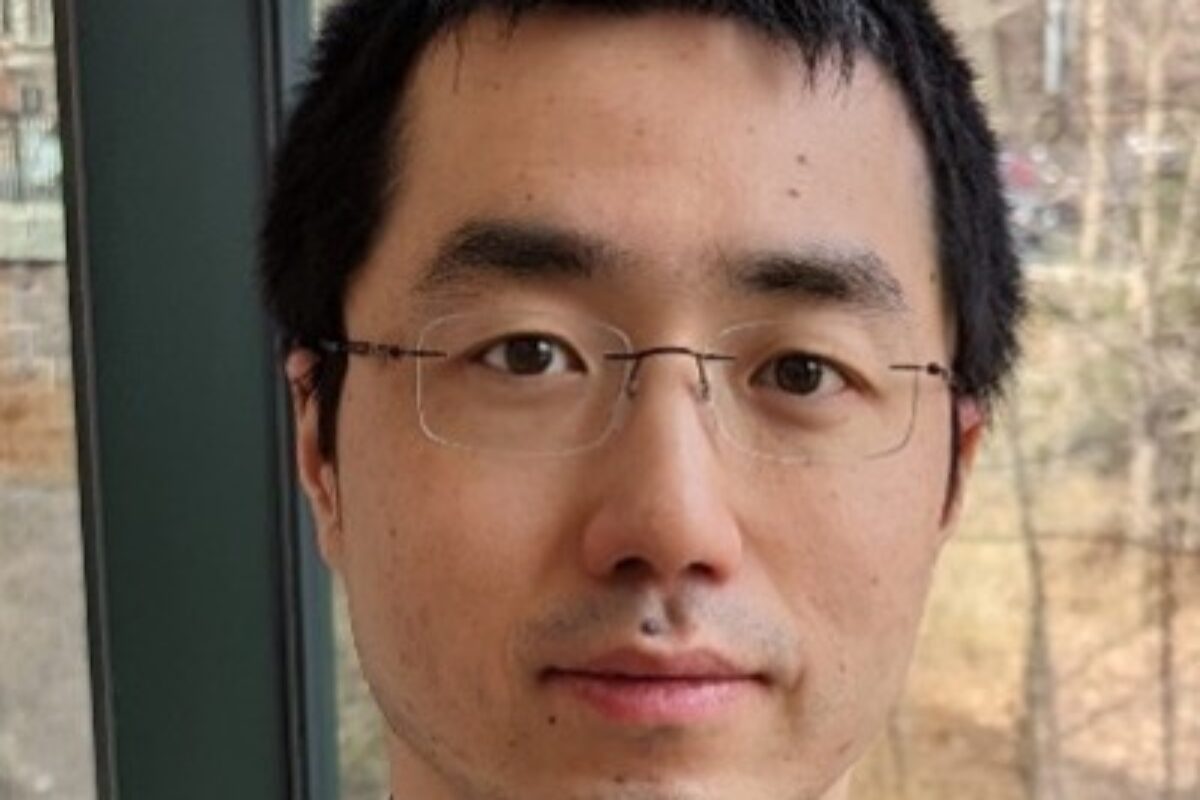
Yanxiang Deng, Ph.D.

  1. Spatially resolved profiling of histone modifications

To investigate the mechanisms underlying spatial organization of different cell types and functions in the tissue context, it is highly desired to examine not only gene expression but also epigenetic underpinnings in a spatially resolved manner to uncover the causative relationship determining what drives tissue organization and function. Despite recent advances in spatial transcriptomics to map gene expression, it has not been possible to determine the underlying epigenetic mechanisms controlling gene expression and tissue development with high spatial resolution. We developed a first-of-its-kind technology called spatial-CUT&Tag for genome-wide profiling of histone modifications pixel by pixel on a frozen tissue section without dissociation. This method resolved spatially distinct and cell-type-specific chromatin modifications in mouse embryonic organogenesis and postnatal brain development. Single-cell epigenomic profiles were derived from the tissue pixels containing single nuclei. Spatial-CUT&Tag adds a new dimension to spatial biology by enabling the mapping of epigenetic regulations broadly implicated in development and disease. In addition, epigenetic drugs are emerging now, so potentially we can develop drugs to target those epigenetic mechanisms. Having the tools to understand the epigenetic origin of different disease states could open up a whole new avenue of therapeutics.

  • Deng, Y., Bartosovic, M., Kukanja, P., Zhang, D., Liu, Y., Su, G., Enninful, A., Bai, Z., Castelo-Branco, G. and Fan, R. Spatial-CUT&Tag: spatially resolved chromatin modification profiling at the cellular level, Science, 375: 681-686 (2022).
  1. Spatially resolved profiling of chromatin accessibility

An ambitious global initiative has been undertaken to map cell types across all human organs. Single-cell sequencing has been critical to this effort, but it is hard to map the location of cell types to the original tissue environment. We developed spatial-ATAC-seq, which for the first time allows for directly observing cell types in a tissue as defined by global epigenetic state. Spatial-ATAC-seq allows us to identify which regions of the chromatin are accessible genome-wide in cells at specific locations in a tissue. This chromatin accessibility is required for genes to be activated, which then provides unique insights on the molecular status of any given cell. Combining the ability to analyze chromatin accessibility with the spatial location of cells is a breakthrough that can improve our understanding of cell identity, cell state and the underlying mechanisms that determine the expression of genes in the development of different tissues or diseases. Profiling mouse embryos using spatial-ATAC-seq delineated tissue-region-specific epigenetic landscapes and identified gene regulators involved in the development of the central nervous system. Mapping the accessible genome in the mouse and human brain revealed the intricate arealization of brain regions. Applying spatial-ATAC-seq to tonsil tissue resolved the spatially distinct organization of immune cell types and states in lymphoid follicles and extrafollicular zones. This technology progresses spatial biology by enabling spatially resolved chromatin accessibility profiling to improve our understanding of cell identity, cell state and cell fate decision in relation to epigenetic underpinnings in development and disease.

  • Deng, Y., Bartosovic, M., Ma, S., Zhang, D., Kukanja, P., Xiao, Y., Su, G., Liu, Y., Qin, X., Rosoklija, B.R, Dwork, A., Mann, J.J., Xu, M.L., Halene, S., Craft, J.E., Leong, W.K., Boldrini, M., Castelo-Branco, G. and Fan, R. Spatial profiling of chromatin accessibility in mouse and human tissues, Nature, 609: 375–383 (2022).
  1. Spatially resolved transcriptomics and proteomics

In multicellular systems, cells do not function in isolation but are strongly influenced by spatial location and surroundings. Spatial gene expression heterogeneity plays an essential role in a range of biological, physiological, and pathological processes. We developed a novel microfluidic platform (DBiT-seq) to deliver molecular barcodes to formaldehyde or FFPE fixed tissue sections in a spatially confined manner, enabling simultaneous barcoding of mRNAs and proteins, and construction of a high-spatial-resolution multi-omics atlas by NGS sequencing. The unique microfluidic in-tissue barcoding technique has enabled high-spatial-resolution mapping of whole transcriptome and tens of proteins at cellular level.

  • Liu, Y#, Yang, M#, Deng, Y#, Su, G, Enninful, A, Guo, C, Tebaldi, T, Zhang, D, Kim, D, Bai, Z, Norris, E, Pan, A, Li, J, Xiao, Y, Halene, S and Fan, R. “High-Spatial-Resolution Multi-Omics Sequencing via Deterministic Barcoding in Tissue”, Cell, 183: 1–17 (2020).

Research Interest

The Deng lab is developing novel technologies for spatial omics to solve challenging biological problems, including cancer and neurodegenerative diseases.

Vikram Paralkar, MD

Research Interest

Research in the Paralkar Lab spans the spectrum from human patient sample studies and mouse models to cutting-edge molecular biology tools, high-throughput sequencing approaches, and novel computational algorithms, all with the goal of gaining insight into how the transcription of coding genes and noncoding ribosomal DNA genes is regulated in hematopoietic stem cells, myeloid progenitors, and in leukemia.

rRNA Transcription in Hematopoiesis and Leukemia

Ribosomal RNA (rRNA) forms the majority of cellular RNA, and its transcription in the nucleolus by RNA Polymerase I from ribosomal DNA (rDNA) repeats accounts for the bulk of all transcription. rRNA transcription rates vary dramatically between different normal cell types in the hematopoietic tree, and leukemic cells have characteristic prominent nucleoli, indicating robust ribosome synthesis.

The rate of ribosome production has far-reaching influence on the fate of the cell, and dictates its size, proliferation, and ability to translate global or specific mRNAs. Little is known however about how rRNA transcription is regulated and fine-tuned across normal and malignant tissues, and whether this regulation can be targeted for leukemia treatment.

The Paralkar Lab has identified that key hematopoietic and leukemic transcription factors bind to rDNA and regulate rRNA transcription, and we are interested in understanding how the binding of cell-type-specific transcription factors regulates the activity of Polymerase I and the transcription of rRNA in normal hematopoiesis, and how this regulation is co-opted in leukemia to drive abundant ribosome biogenesis.

Stemness and Differentiation in Hematopoiesis and Leukemia

Normal hematopoiesis requires an intricate balance in the bone marrow between the ability of stem cells to maintain themselves for decades of life while producing billions of mature blood cells every day. This balance is maintained by the combinatorial activity of transcription factors and chromatin proteins that dictate the transcription of coding gene networks instructing fate choice decisions. Several of the critical factors involved in these decisions are mutated in acute and chronic leukemias, and their mutations tip the equilibrium in the bone marrow towards accumulation of aberrant progenitor populations.

The Paralkar Lab is interested in gaining a detailed mechanistic understanding of how chromatin proteins regulate the stemness-differentiation balance, and how mutations in them produce malignancy.

Bioinformatic Pipelines for Genetics and Epigenetics

Current bioinformatic pipelines for high-throughput studies like whole genome sequencing, RNA-seq, ChIP-seq, and single cell RNA-seq are limited in their ability to map repetitive elements of the genome like ribosomal DNA. Such loci therefore tend to be ignored in genome wide analyses. Given that rRNA accounts for the bulk of the transcriptional output of the cell, the inability to map datasets to rDNA has historically been a major limitation, and has created a significant knowledge gap in our understanding of the most abundant RNA in the cell.

The Paralkar Lab has developed customized genomes and computational pipelines to map datasets to rDNA, and we are interested in developing advanced tools to map and interpret the genetic and epigenetic profiles of rDNA in normal and malignant cells.

Selected Publications

George SS, Pimkin M, Paralkar VR: Customized genomes for human and mouse ribosomal DNA mapping. BioRxiv Nov 2022.

Antony C, George SS, Blum J, Somers P, Thorsheim CL, Wu-Corts DJ, Ai Y, Gao L, Lv K, Tremblay MG, Moss T, Tan K, Wilusz JE, Ganley ARD, Pimkin M, Paralkar VR: Control of ribosomal RNA synthesis by hematopoietic transcription factors. Molecular Cell 82(20): 3826-3839, Oct 2022.

Lv K, Gong C, Antony C, Han X, Ren J, Donaghy R, Cheng Y, Pellegrino S, Warren AJ, Paralkar VR, Tong W: HectD1 controls hematopoietic stem cell regeneration by coordinating ribosome assembly and protein synthesis. Cell Stem Cell 28: 1-16, Jul 2021.

Xu P, Palmer LE, Lechauve C, Zhao G, Yao Y, Luan J, Vourekas A, Tan H, Peng J, Scheutz JD, Mourelatos Z, Wu G, Weiss MJ, Paralkar VR: Regulation of gene expression by miR-144/451 during mouse erythropoiesis. Blood 133(23): 2518-2528, Jun 2019.

Traxler EA, Thom CS, Yao Y, Paralkar V, Weiss MJ: Non-specific inhibition of erythropoiesis by short hairpin RNAs. Blood 131(24): 2733-2736, Jun 2018.

Paralkar VR, Taborda CC, Huang P, Yao Y, Kossenkov AV, Prasad R, Luan J, Davies JO, Hughes JR, Hardison RC, Blobel GA, Weiss MJ: Unlinking an lncRNA from Its Associated cis Element. Molecular Cell 62(1): 104-10, Apr 2016.

Paralkar VR, Mishra T, Luan J, Yao Y, Kossenkov AV, Anderson SM, Dunagin M, Pimkin M, Gore M, Sun D, Konuthula N, Raj A, An X, Mohandas N, Bodine DM, Hardison RC, Weiss MJ: Lineage and species-specific long noncoding RNAs during erythro-megakaryocytic development. Blood 123(12): 1927-37, Mar 2014.

Mustafa A. Mir, Ph.D.

1. Label-free blood screening instruments (Masters work). Whole blood analysis is a critical and familiar part of clinical workflows. Widely used clinical blood cytometers are generally expensive and bulky, require proprietary reagents, and provide limited quantitative information on cell morphology. When these automated cytometers do find abnormalities, manual examination of a stained blood smear using light microscopy is required for further diagnosis. This process is costly, slow, and not quantitative. In my early graduate career, working Prof. Gabriel Popescu, I set out to address these issues by utilizing Quantitative Phase Imaging (QPI). In QPI the phase shift (optical delay) of light passing through a sample is measured with sub-nanometer accuracy using interferometry providing high resolution topographic and tomographic data (Mir et al, Prog. Optics, 2012). I started by building a new QPI instrument utilizing commercial CD-ROM technology (Mir et al., Opt. Express., 2009) with the goal of developing a low-cost platform for an automated point-of-care blood screening tool. A comparison of results on patient samples with those from a clinical cytometer shows excellent agreement (Mir et al., J. Biomed. Opt., 2010 & Biomed. Opt. Exp., 2011) for clinical parameters on red blood cells. Additionally, my approach provides quantitative single cell morphological data, uses 2 orders of magnitude lower input sample, requires no reagents other than whole blood, and significantly lowers instrument cost. Collectively, these studies established that QPI based technologies are more sensitive than currently used clinical whole blood analyzers for common parameters used to characterize erythrocytes and also provide additional information that could lead to earlier diagnosis of certain diseases. 

a) M. Mir, B. Bhaduri, R. Wang, R. Zhu, G. Popescu, Chapter 3 – Quantitative Phase Imaging, Progress in Optics, Ed: W. Emil, Elsevier, Volume 57, pp. 133-217 (2012) 

b) M. Mir, Z. Wang, K. Tangella and G. Popescu, Diffraction Phase Cytometry: Blood on a CD-ROM, Optics Express, 17 (4), 2579-2585 (2009) 

c) M. Mir, H. Ding, Z. Wang, J. Reedy, K. Tangella and G. Popescu, Blood Screening using Diffraction Phase Cytometry, Journal of Biomedical Optics, 15 (2), 027016 (2010) 

d) M. Mir, K. Tangella and G. Popescu, Blood Testing at the Single Cell Level using Quantitative Phase and Amplitude Microscopy, Biomedical Optics Express, 2 (12), 3259-3266. (2011) 

2. Non-perturbative, quantitative, and label-free live imaging spanning broad spatio-temporal scales (PhD work). A major challenge in the live imaging of cells and tissues is the need to non-destructively capture data at a broad range of temporal and spatial scales. High temporal and spatial resolution data is required to measure dynamics and morphological changes at the sub-cellular level, at sub-micron and millisecond scales, while at the same time placing this data in the context of the population (e.g. whole tissue) at scales of millimeters and days. In Prof. Gabriel Popescu’s lab we developed new quantitative phase imaging (QPI) hardware and analytical tools which provide quantitative information on morphology, mass density, and dynamics while spanning this broad range of spatial-temporal scales. Critically, these new microscopes use low-power white-light illumination and are sensitive to the endogenous optical properties of the sample, circumventing challenges associated with fluorescence microscopy such as photobleaching and phototoxicity (at the expense of molecular specificity). One of our instruments, known as the Spatial Light Interference Microscope (Wang et al, Opt. Express, 2011), for which I developed control hardware and software, has also been successfully commercialized (phioptics.com). I used these new tools to 1) Characterize single cell mass growth in a cell-cycle dependent manner, overturning widely used models of constant exponential growth (Mir et al, PNAS, 2011); 2) Measure the effects of estrogen and estrogen agonists on the growth and dynamics of estrogen sensitive breast cancer cells, showing that estrogen blockers lead to slower growing cells with lower cell masses (Mir et al, PLoS One, 2014); 3) Quantify neuronal network formation in human stem cell derived neurons and showed correlations between single cell mass growth, transport within the forming network, and the emergence of self-organization of the developing network (Mir et al, Sci. Rep., 2014). These studies have had a major impact on the rapid growth of the field of Quantitative Phase Imaging and its adoption by life scientists which is best illustrated by the increasing presence of dedicated QPI instruments in core facilities and laboratories around the world. 

a) Z. Wang, L. Millet, M. Mir, H. Ding, S. Unarunotai, J. Rogers, M. Gillette, and G. Popescu, Spatial Light Interference Microscopy, Optics Express, 19 (2), 1016-1026 (2011) 

b) M. Mir, Z. Wang, Z. Shen, M. Bednarz, R. Bashir, I. Golding and G. Popescu*, Optical Measurement of Cell Cycle Dependent Growth, Proceedings of the National Academies of Sciences (PNAS), 108 (32), 13124-13129 (2011) 

c) M. Mir, A. Bergamaschi, B.S. Katzenellenbogen and G. Popescu, Highly sensitive quantitative imaging for single cancer cell growth kinetics and drug response, PLoS ONE, 9 (2), e89000 (2014) 

d) M. Mir, T. Kim, A. Majumder, M. Xiang, R. Wang, S. C. Liu, M. U. Gillette, S. Stice and G. Popescu, Label-Free Characterization of Emerging Human Neuronal Networks” Scientific Reports, 4, 4434 (2014) 

3. Computational Imaging (PhD and Postdoc work). Live-cell microscopy involves harsh tradeoffs between spatial resolution, temporal resolution, and sensitivity. In all optical microscopes spatial resolution is limited by the diffraction limit of light. Additionally, in the case of fluorescence microscopy, photo-bleaching and photo-toxicity present significant barriers for high-speed 3D imaging. Over the past two decades the field of compressed sensing has emerged to take advantage of an intrinsic property of natural images known as sparsity. Sparsity can be exploited to reconstruct high resolution images from lower resolution data (time or space) provided this data is acquired in an appropriate manner. Such computational imaging approaches, integrate hardware design and computational algorithms to boost spatial and temporal imaging resolution. During my graduate work, I developed a 3D deconvolution method (with Dr. D. Babacan) that exploits the sparsity of quantitative phase images to provide super-resolution 3D tomograms of living cells in a completely label-free manner (Mir et al., PLoS ONE, 2012). Using this approach I was able to resolve and characterize coiled and helical sub-cellular structures in E. coli which were previously only visible using fluorescence based super-resolution methods. We then further refined this approach by including a near-complete physical model of our optical system in a technique known as White-Light Diffraction Tomography (Kim et al., Nat. Photonics, 2014) which provides quantitative 3D tomograms of live cells with sub-cellular resolution. During my post-doctoral work, I led a project (senior author) to develop a generalizable compressed sensing scheme which provides a 5-10 fold reduction of light exposure and acquisition time in 3D fluorescence microscopy (Woringer et al., Opt. Express 2017). Collectively these works have demonstrated novel computational imaging approaches to improve the temporal and spatial resolution of light-microscopes and decrease photo-bleaching and photo-toxicity in 3D fluorescence imaging. 

a) M. Mir, D. Babacan, M. Bednarz, I. Golding and G. Popescu, Three Dimensional Deconvolution Spatial Light Interference Tomography for studying Subcellular Structure in E. coli , PLoS ONE, 7 (6), e38916 (2012) 

b) T. Kim, R. Zhou, M. Mir, S. D. Babacan, P. S. Carney, L. L. Goddard, and G. Popescu, White-light diffraction tomography of unlabelled live cells, Nature Photonics, (2014) 

c) M. Woringer, X. Darzacq, C. Zimmer, and M. Mir, Faster and less phototoxic 3D fluorescence microscopy using a versatile compressed sensing scheme, Optics Express, 25(12), 13668-13683, (2017) 

4. Single molecule tracking and high-resolution 4D imaging in live embryos (Postdoc work). The application of single-molecule tracking to study transcription factor dynamics has shed significant insight on their search and DNA-binding kinetics over the past decade in cell culture models. At the start of my postdoc the technology to perform such measurements in live embryos did not exist. I overcame this problem by building a customized lattice light-sheet microscope to acquire the first data on single-molecule kinetics of proteins within the nuclei of living animal embryos (demonstrated in Mice, C. elegans, and Drosophila, Mir et al., Methods Mol Biol., 2018). In addition to enabling single molecule tracking in embryos, lattice light-sheet microscopy also provides high resolution 4D imaging over large fields-of-views and extended periods of times. These unique imaging capabilities provided opportunities for several collaborative efforts centered on questions of nuclear organization and transcription regulation (these collaborative works are in addition to my primary focus on studying transcription factor binding in Drosophila embryos as described below). Notably, these efforts include 1) Revealing the role of phase separation in the formation of heterochromatin domains in collaboration with Gary Karpen’s group (Strom et al, Nature 2017), 2) Characterizing the role of intrinsically disordered regions in replication initiation factor loading in collaboration with Michael Botchan’s group (Parker et al., eLife 2019), 3) Developing capabilities to label non-repetitive genomic loci with Cas9 in collaboration with Ahmet Yildiz’s group (Qin et al, 2017) and 4) Using single molecule-imaging in C. elegans embryos to study the regulation of RNA polymerase II recruitment to chromatin during X-chromosome dosage compensation in collaboration with Barbara Meyer’s group (in preparation). These collaborative efforts have provided unique biophysical insights on nuclear organization, chromatin biology, and transcription regulation and have helped mature the technologies of single molecule imaging in live embryos and software to analyze large high-dimensional imaging datasets. 

a) M. Mir, A. Reimer, M. Stadler, A. Tangara, A. S. Hansen, D. Hockemeyer, M. B. Eisen, H. Garcia, X. Darzacq*, Chapter 32 – Single Molecule Imaging in Live Embryos using Lattice Light-Sheet Microscopy, Methods in Molecular Biology, Nanoscale Imaging: Methods and Protocols, Volume 1814, Ed: Y. L. Lyubchenko, Springer, pp 541-559 (2018) 

b) A. R. Strom, A. V. Emelyanov, M. Mir, D. V. Fyodorov, X. Darzacq, and G. Karpen, Phase separation drives heterochromatin domain formation, Nature, doi:10.1038/nature22989 (2017) 

c) M. W. Parker, M. Bell, M. Mir, J. A. Kao, X. Darzacq, M. R. Botchan, J. M. Berger, A new class of disordered elements controls DNA replication through initiator self-assembly, eLife 8:e48562, (2019) Download PDF 

d) P. Qin, M. Parlak, C. Kuscu, J. Bandaria, M. Mir, K. Szlachta, R. Singh, X. Darzacq, A. Yildiz, and M. Adli, Live cell imaging of low- and non- repetitive chromosome loci using CRISPR-Cas9, Nature Communications, 8, 14725, (2017) 

5. High concentration multi-factor hubs accentuate transcription factor binding and gene activation (Postdoc work). During my postdoc I became fascinated with linking transcription regulation at the molecular scale to cell-fate patterning during embryonic development. Central to this question is the biophysical problem of how transcription factors find their specific genomic targets in the crowded nuclear milieu. I utilized single molecule imaging to study the anteroposterior morphogen gradient formed by the transcription factor Bicoid in Drosophila melanogaster embryos. Bicoid has long been a model for classical cooperative binding, in which protein-protein interactions stabilize its interaction with target sites. However, when I analyzed Bicoid dynamics in nuclei along its anterior to posterior gradient, I saw no evidence for the concentration-dependent binding off-rates this model predicts. My data instead revealed that Bicoid binding occurs in high-local concentration hubs, which are dependent on the presence of a maternally deposited pioneer factor, Zelda. These multi-factor hubs increase the frequency of Bicoid binding events and thus result in higher Bicoid time-averaged occupancy without directly stabilizing its interactions with DNA (Mir et al, Genes Dev. 2017). Remarkably, these hubs themselves are highly dynamic, forming and melting on the order of seconds. By combining single-molecule tracking, high-speed 3D imaging, and live-visualization of nascent transcription I showed that Zelda-Bicoid hubs drive enrichment of transcription factors at their target loci through transient but preferential interactions (Mir et al, eLife, 2018) which then result in bursts of transcriptional activity (unpublished). Recently I have shown that hub formation is dependent on interactions between the disordered protein domains of hub components and independent of DNA binding (unpublished). These discoveries have come at a time when there is great interest in the role of disordered proteins in driving nuclear organization, particularly in the context of liquid-liquid phase separation. However, my work illustrates that disordered protein interactions can lead to dynamic compartmentalization distinct from phase separation, emphasizing that we have barely begun to understand the myriad biophysical forces that are at play (Mir et al, Development, 2019; McSwiggen et al., Genes Dev 2019). 

a) M. Mir, A. Reimer, J. E. Haines, X.Y. Li, M. Stadler, H. Garcia, M. B. Eisen, X. Darzacq, Dense Bicoid hubs accentuate binding along the morphogen gradient, Genes Dev., 31, 1784-1794 (2017) 

b) M. Mir, M. R. Stadler, S.A. Ortiz, X. Darzacq, M. B. Eisen, Dynamic Multifactor hubs transiently interact with sites of active transcription in Drosophila embryos, eLife, 7:e40497, (2018) 

c) M. Mir, W. Bickmore, E. E. M. Furlong, G. J. Narlikar, Chromatin topology, condensates, and gene regulation: shifting paradigms or just a phase? Development, 146,dev182766 (2019) 

d) D. T. McSwiggen, M. Mir, X. Darzacq, R. Tjian, Evaluating phase separation in live cells: diagnosis, caveats, and functional consequences, Genes Dev, (2019) 

Research Interest

Gene expression is regulated by a complex choreography of highly dynamic events, including the binding of transcription factors to non-coding regulatory regions of the genome, regulation of chromatin topology, and the assembly of large macromolecular complexes, all of which occur in the crowded nuclear environment. Our understanding of these dynamic processes has largely been driven by approaches that provide population averaged and static snapshots which have delivered remarkable insights, but are inherently ill suited for elucidating processes that vary greatly in space and time.  Comprehending the mechanisms that regulate gene expression, and the role of nuclear organization in this regulation, requires technological and theoretical approaches that bridge spatial scales from molecular to organismal and temporal scales from milliseconds to days. We develop and utilize high-resolution microscopy methods which allows us to probe this vast range of spatial and temporal scales within living embryos. For example, we acquire high-speed volumetric data to quantify chromatin dynamics, multi-color datasets  to study the interaction and distribution of protein domains associated with gene activation or repression as cell fates are determined in young embryos, and use single-molecule localization techniques to quantify the kinetics of individual transcription factors as they whizz around the nucleoplasm searching for and binding to their genomic targets. The goal of our lab is to use these advanced imaging technologies in combination with biophysical modelling, genomics, and gene editing to comprehend and manipulate the interplay between nuclear organization, transcription regulation, and gene expression patterns during cell-fate determination.

Melike Lakadamyali, Ph.D.

  1. Developed versatile methods for quantitative, multiplexed imaging of molecular complexes using super-resolution microscopy:Proteins in cells assemble into nanoscale complexes in order to carry out a specific function. The spatial organization and stoichiometry of proteins within these nanoscopic functional units is highly important for maintaining a cell’s healthy physiology. Changes in nanoscale organization of protein complexes, for example a change from monomeric to oligomeric stoichiometry, often triggers disease states. However, visualizing many proteins simultaneously and quantifying protein copy number with high spatial resolution is highly challenging. Super-resolution methods hold promise for overcoming this hurdle, however, the complex photophysics of fluorophores limits both multi-color imaging capabilities and the ability to extract quantitative information. Lakadamyali lab has been developing new methods to overcome these challenges. To address the limitations in simultaneous, high-throughput, multi-color super-resolution imaging, we recently combined DNA-Paint with excitation multiplexing to demonstrate the initial proof of concept for fm-DNA-Paint. Further, to address the challenges with quantification of protein copy number at the nanoscale level, we built calibration standards based on DNA origami. Overall, these methods begin to overcome some of the main challenges associated to super-resolution microscopy making it a multiplexed and quantitative tool.


  1. “Quantifying protein copy number in super-resolution using an imaging invariant calibration”,F.C. Zanacchi, C. Manzo, R. Magrassi, N.D. Derr,M. Lakadamyali, Biophysical Journal, 116, 2195-2203 (2019)
  2. “Excitation-multiplexed multicolor super-resolution imaging with fm-STORM and fm-DNA-Paint” P.A. Gómez-García, E.T. Garbacik, M.F. Garcia-Parajo,M. Lakadamyali,PNAS, 115, 12991-12996 (2018)
  3. “DNA Origami: Versatile super-resolution calibration standard for quantifying protein copy number” F. Cella Zanacchi, C. Manzo, A. Sandoval Alvarez, N, Derr,M. Lakadamyali,Nature Methods, doi:10.1038/nmeth.4342 (2017)
  4. “Single molecule evaluation of fluorescent protein photoactivation efficiency using anin vivonanotemplate”, N. Durisic, L. L. Cuervo, A. S. Álvarez, J.Borbely, M. LakadamyaliNature Methods, 11, 156-162 (2014)
  1. Determined a novel nanoscale organization of chromatin:An important goal of the Lakadamyali lab is to reconcile the epigenomic and microscopic views of chromatin organization and determine how chromatin structure regulates gene function. Nuclear organization of the chromatin fiber spans many length-scales and while the nanoscale level organization plays a key role in regulating gene expression, this organization is impossible to visualize using conventional microscopy methods. Lakadamyali lab has been using and further developing quantitative super-resolution microscopy methods to overcome this limitation. We visualized and estimated the number of nucleosomes along the chromatin fiber of different cells at nanoscale resolutionWe discovered that nucleosomes are assembled in heterogeneous groups of varying sizes, which we termed “clutches”. Remarkably, the median number of nucleosomes and their packing density inside clutches highly correlated with cellular state, such that clutch size correlates with gene expression and pluripotency grade of iPSCsAdditionally, nanoscale chromatin organization is aberrantly remodeled in disease states.
  2. “Aberrant chromatin reorganization in cells from diseased fibrous connective tissue in response to altered chemomechanical cues”. S. Heo, S. Thakur, X. Chen, C. Loebel, B. Xia, R. McBeath, J.A. Burdick, V.B. Shenoy, R.L. Mauck, M. LakadamyaliNature Biomedical Engineering, in press (2022)

b.     “Two-Parameter mobility assessments discriminate diverse regulatory factor behaviors in chromatin”, J. Lerner, P.A. Gomez-Garcia, R. McCarthy, Z. Liu, M. Lakadamyali, K. S. Zaret, Molecular Cell, doi: https://doi.org/10.1016/j.molcel.2020.05.036, (2020)

  1. “Super-resolution microscopy reveals how histone tail acetylation affects DNA compaction within nucleosomes in vivo”, J. Otterstrom, A.C. Garcia, C. Vicario, P.A. Gomez-Garcia, M.P. Cosma, M. LakadamyaliNucleic Acids Research,  doi: 10.1093/nar/gkz593 (2019)
  2. “Chromatin fibers are formed by heterogeneous groups of nucleosomes in vivo”, M. A. Ricci, C. Manzo, M. Garcia-Parajo, M. Lakadamyali, M. P. Cosma, (co-senior authors), Cell160, 1145-1158 (2015)

3. Developed cutting-edge methods for studying intracellular trafficking: One of the overarching goals of Lakadamyali lab is to determine how molecular motors coordinate to transport vesicles and organelles in the complex cellular environment. To achieve this goal, we developed an “all-optical correlative imaging method” that combines single particle tracking in living cells with super-resolution microscopy of the same cell after fixation. Super-resolution microscopy can reveal the details of cellular architecture with exquisite spatial resolution; however, the current methods cannot capture fast dynamic processes (millisecond time scale) due to limited temporal resolution. With the correlative approach, we overcame this limitation and for the first time related cargo transport dynamics to the organization of the microtubule cytoskeleton in the cellular context. By mapping cargo trajectories to individual microtubules, we studied how the local cytoskeletal architecture regulates vesicle trafficking. We found that the cytoskeleton acts as a selective filter to vesicles that are comparable in size to or larger than the mesh size of the microtubule network leading to their pausing. We further showed that lysosomal and autophagosomal compartments are selectively enriched on specific microtubule tracks defined by post-translational modifications in order to enhance their fusion and regulate autophagy. Overall, we have developed powerful methods that reveal how the 3D local cytoskeletal architecture and the microtubule post-translational modifications combine to regulate, opening a new window into studying vesicle-cytoskeleton interactions in the physiological as well as disease contexts. We have recently expanded these studies to visualizing the distribution of microtubule associated proteins like tau under physiological and pathological conditions.

  1. “Tau forms oligomeric complexes on microtubules that are distinct from pathological oligomers”, M.T. Gyparaki, A. Arab, E.M. Sorokina, A.N. Santiago-Ruiz, C.H. Bohrer, J. Xiao, M. Lakadamyali, PNAS, 118 (19) e2021461118 (2021)
  2.  Detyrosinated microtubules spatially constrain lysosomes facilitating lysosome-autophagosome fusion” N. Mohan, I. V. Verdeny, A. S. Alvarez, E. Sorokina, M. Lakadamyali, Journal of Cell Biology, doi: 10.1083/jcb.201807124, (2018)
  3. “3D Motion of Vesicles Along Microtubules Helps Them to Circumvent Obstacles in Cells”, I. V. Vilanova, F. Wehnekamp, N. Mohan, A. S. Alvarez, J. S. Borbely, J. Otterstrom, D. Lamb, M. LakadamyaliJournal of Cell Science, doi: 10.1242/jcs.201178, (2017)
  4. Correlative live-cell and superresolution microscopy reveals cargo transport dynamics at microtubule intersections”, Š. Bálint, I. V. Verdeny, A. S. Álvarez, M. LakadamyaliPNAS1103375-3380 (2013) (cover page illustration)

Research Interest

The overarching goal of the Lakadamyali lab is to understand the molecular mechanisms that regulate sub-cellular organization and the significance of this organization on cell function. Cells are highly compartmentalized: the sub-cellular positioning of organelles, nucleic acids and proteins are spatially and temporally coordinated to ensure that biochemical reactions take place at the right place and time. Dr. Lakadamyali’s program has three major focus areas that seek to advance our understanding of sub-cellular organization. First, she develops advanced microscopy methods that provide the technological advances necessary to visualize the spatial organization of cellular machinery with near molecular spatial resolution, including quantitative tools for measuring protein stoichiometry and multiplexed, high-throughput imaging methods. Second, she seeks to determine how the microtubule cytoskeleton and motors regulate transport and positioning of organelles within the cytoplasm and the functional consequences of disrupting proper organelle organization. As part of this work, her lab has recently expanded their focus to studying microtubule associated protein tau and its aggregation in neurological diseases. Third, she seeks to understand how the spatial organization of chromatin within the nucleus regulates gene activity. These three areas integrate synergistically to move forward our understanding of how sub-cellular organization emerges and impacts cell physiology and pathology.

Maya Capelson, Ph.D.

Contact Information

The Perelman School of Medicine at the University of Pennsylvania
Department of Cell and Developmental Biology
9-101 Smilow Center for Translational Research
3400 Civic Center Blvd
Philadelphia, PA 19104-6059
Office: 215-898-0550
Lab: 215-573-7548
capelson@pennmedicine.upenn.edu

Robert Babak Faryabi, Ph.D.

Deciphering Mechanisms of Genome Mis-folding In Cancer

Our lab deploys data-rich experimental techniques to elucidate the role of genome mis-folding in controlling oncogenic gene expression programs. Specifically, we are interested in moving beyond the status quo to understand how oncogenic subversion of lineage-determining transcription factors set topology of cancer genome. To tackle this question, we combine genomics and super-resolution imaging and focus on investigating molecular mechanisms of genome mis-folding in breast and blood cancers. These mechanistic studies aim to identify precise epigenetic vulnerabilities of cancer cells and guide treatments disrupting cancer cells’ transcriptional addiction.

Representative Publication:

Oncogenic Notch Promotes Long-Range Regulatory Interactions Within Hyperconnected 3D Cliques. Petrovic J*, Zhou Y*, Fasolino M, Goldman N, Schwartz GW, Mumbach MR, Nguyen SC, Rome KS, Sela Y, Zapataro Z, Blacklow SC, Kruhlak MJ, Shi J, Aster JC, Joyce EF, Little SC, Vahedi G, Pear WS, Faryabi RB Molecular Cell. 2019;73(6):1174-90 e12

Determining Epigenetic Mechanisms Of Resistance To Targeted Therapies

Targeting oncogenic drivers of cancers commonly leads to drug resistance. Mechanisms of acquiring resistance to oncology drugs mostly remain unknown, partly due to the limitations of population-based assays in elucidating heterogeneity of drug-naive and complexity of drug-induced tumor evolution. Using single-cell genomics and imaging, we study how heterogeneity and plasticity of transcriptional dependencies confer resistance to targeted therapeutics such as Notch inhibitors.

Representative Publication

TooManyCells Identifies And Visualizes Relationships Of Single-cell Clades. Schwartz GW, Zhou Y, Petrovic J, Fasolino M, Xu L, Shaffer SM, Pear WS, Vahedi G, Faryabi RB Nature Methods, 2020; 17: 405-413

Innovating Computational Methods To Enable Cancer Discovery

Our lab innovates statistical and machine learning approaches to accelerate discovery of novel therapeutics and biomarkers by elucidating complexity and heterogeneity of tumors. Recently, we have developed a computational ecosystem for mapping molecular and spatial heterogeneity in tumors. As part of the Center for Personalized Diagnostics, we also mine cancer patient genotypic/phenotypic data to improve patient health. patient health.

Representative Publication:

Classes of ITD Predict Outcomes in AML Patients Treated With FLT3 Inhibitors. Schwartz GW, Manning B, Zhou Y, Velu P, Bigdeli A, Astles R, Lehman AW, Morrissette JJD, Perl AE, Li M, Carroll M, Faryabi RB Clinical Cancer Research. 2019;25(2):573-83

Research Interest

Cancer is typically considered a genetic disease. However, recent progress in our understanding of epigenetic aberrations in cancer has challenged this view. Overarching goal of our lab is to understand epigenetic mechanisms of transcriptional addiction in cancer and exploit this information to advance cancer therapeutics.

To pursue this objective, we use cutting-edge chromatin conformation capture, high-content imaging, single-cell epigenomics, functional genomics, and combine these technologies with our expertise in computational sciences to systematically explore: i) how epigenetic control of gene expression is disrupted in cancer, ii) why transcriptional addiction can develop, and iii) how heterogeneity and plasticity of transcriptional dependencies enable drug resistance.

Liling Wan, Ph.D.

Molecular link between histone acetylation and oncogenic gene activation Histone acetylation is a chromatin mark generally associated with gene activation, yet the molecular mechanisms underlying this correlative relationship remain incompletely understood. I led a collaborative study in which we identified a novel ‘reader’ for histone acetylation named ENL. We showed in leukemia cells that ENL interacts with histone acetylation via the well-conserved YEATS domain, and in so doing, helps to recruit and stabilize its associated transcriptional machinery to drive transcription of leukemogenic genes. By determining the structure of ENL in complex with an acetylated histone peptide, we and our collaborators demonstrated that disrupting the reader function reduced chromatin recruitment of ENL-associated transcriptional machinery and resulted in suppression of oncogenic programs. Furthermore, blocking the functionality of ENL sensitized leukaemia cells to inhibitors that target another distinct class of histone acetylation readers, the BET proteins, thus highlighting the crosstalk between epigenetic readers and potential benefit of combinatorial therapies. Our work established ENL as a missing molecular link between histone acetylation and gene activation critical for leukemia malignant state, and has inspired following studies investigating other YEATS domain-containing proteins as a new class of chromatin ‘readers’ in a broad range of human cancers. In addition to bringing novel insights into our basic understanding of chromatin regulation, this work also provides mechanistic guidance and structural basis for ongoing drug development to target chromatin reading activity of ENL in aggressive leukemias.

Wan L#, Wen H#, Li Y#, Lyu J, Xi Y, Hoshii T, Joseph JK, Wang X, Loh YE, Erb MA, Souza AL, Bradner JE, Shen L, Li W, Li H*, Allis CD*, Armstrong SA*, Shi X*. ENL Links Histone Acetylation to Oncogenic Gene Activation in Leukemias. Nature 2017 Mar 9;543(7644):265-269. (#Equal contribution). PMC5372383
Li Y*, Sabari BR*, Panchenko T*, Wen H, Zhao D, Guan H, Wan L, Huang H, Tang Z, Zhao Y, Roeder RG, Shi X, Allis CD, Li H. Molecular Coupling of Histone Crotonylation and Active Transcription by AF9 YEATS Domain. Mol Cell2016 Apr 21;62(2):181-93. (*Equal contribution) PMC4841940

New type of gain-of-function mutations in chromatin readers Recognition of modified histones by ‘reader’ proteins constitutes a key mechanism mediating the function of histone modifications, yet the mechanisms by which their dysregulation contributes to diseases remain poorly understood. Recurrent, hotspot mutations in the acetylation-reading domain (YEATS domain) of ENL were recently found in Wilms’ tumor, the most common type of pediatric kidney cancer. Whether and how these mutations cause the disease remained unknown. Our current work shows that these mutations confer ENL gain of function in driving abnormal gene expression implicated in cancer. Unexpectedly, these mutations promoted ENL self-association, resulting in the formation of discrete nuclear puncta that are characteristic of biomolecular condensates, a newly recognized form of protein assembly that often involves weak, multivalent molecular interactions and commonly underlies the formation of membrane-less organelles. We demonstrated that such a property drives ‘self-reinforced’ chromatin targeting of mutant ENL protein and associated transcriptional machinery, thus enforcing active transcription from target loci. Aberrant gene control driven by ENL mutations, in turn, perturbs developmental programs and derails normal cell fate to a path towards tumorigenesis. This work is a remarkable demonstration of how mistakes in chromatin reader-mediated process can act as a driving force for tumor formation. These mutations represent a new class of oncogenic mutations which impair cell fate through promoting self-association and reinforcing chromatin targeting.

Wan L#, Chong S, Fan X, Liang A, Cui X, Gates L, Carroll TS, Li Y, Feng L, Chen G, Wang S, Ortiz MV, Daley S, Wang X, Xuan H, Kentsis A, Muir TW, Roeder RG, Li H, Li W, Tjian R, Wen H#, Allis CD#. Impaired Cell Fate through Gain-of-function Mutations in a Chromatin Reader. Nature 2019 in press (#co-corresponding)

Targeting chromatin readers as cancer therapies My postdoctoral work suggested that the displacement of histone acetylation reader ENL from chromatin may be a promising epigenetic therapy, alone or in combination with BET inhibitors, for aggressive leukemia. I have contributed to the development of peptidomimetic and small molecule inhibitors targeting the YEATS domain protein family. The ultimate goal of these and other ongoing efforts is to develop chemical probes targeting the ‘reading’ activity of ENL and other family members as valuable research tools and potential therapeutic agents.

Li X*, Li XM*, Jiang Y, Liu Z, Cui Y, Fung K, van der Beelen S, Tian G, Wan L, Shi X, Allis CD, Li H, Li Y#, Li X#. Structure-guided Development of YEATS Domain Inhibitors by Targeting π-π-π Stacking. Nat Chem Biol. 2018 Dec;14(12):1140-1149. (*Equal contribution) PMC6503841

Molecular mechanisms of cancer metastasis Metastasis accounts for > 90% cancer-related deaths and yet is the most poorly understood aspect of cancer biology. I have contributed to studies in which we identified and characterized new molecular mechanisms for cancer metastasis. My graduate work focused on Metadherin (MTDH), a novel cancer gene identified by our group to be prevalently amplified in breast cancer and strongly associated with a high risk of metastasis and poor prognosis. What drives the strong selection of MTDH in primary tumors was unclear. By generating genetically engineered mouse models, we provided first evidence supporting an essential role of MTDH in the initiation and metastasis of diverse subtypes of breast cancer. We further showed that MTDH regulates the expansion and activity of cancer stem cells through working with its binding partner SND1. By determining the atomic structure of the complex via collaboration, we demonstrated that disrupting the complex impairs breast cancer development and metastasis in vivo. Our work establishes MTDH and SND1 as critical regulators of cancer development and provides mechanistic guidance for ongoing drug development efforts to target this complex as cancer therapy. More broadly, this work provides crucial experimental support for the emerging concept that metastatic potential could be conferred by early oncogenic events that possess additional metastasis-promoting function and advances our understanding of the origin of metastatic traits.

Wan L, Lu X, Yuan S, Wei Y, Guo F, Shen M, Yuan M, Chakrabarti R, Hua Y, Smith HA, Blanco MA, Chekmareva M, Wu H, Bronson RT, Haffty BG, Xing Y, Kang Y. MTDH-SND1 Interaction Is Crucial for Expansion and Activity of Tumor-Initiating Cells in Diverse Oncogene- and Carcinogen-Induced Mammary Tumors. Cancer Cell 2014 Jul 14;26(1):92-105. PMC4101059
Wan L, Pantel K, Kang Y. Tumor Metastasis: Moving New Biological Insights into the Clinic. Nature Medicine 2013 Nov;19(11):1450-64. PMID: 24202397
Guo F#, Wan L#, Zheng A, Stanevich V, Wei Y, Satyshur KA, Shen M, Lee W, Kang Y, Xing Y. Structural Insights into the Tumor-Promoting Function of the MTDH-SND1 Complex. Cell Reports 2014 Sep 25;8(6):1704-13.  (#Equal contribution). PMC4309369
Wan L, Hu G, Wei Y, Yuan M, Bronson RT, Yang Q, Siddiqui J, Pienta KJ, Kang Y. Genetic Ablation of Metadherin Inhibits Autochthonous Prostate Cancer Progression and Metastasis. Cancer Research 2014 Sep 15;74(18):5336-47. PMC4167565
Kang Y, Xing Y, Wan L, Guo F. Use of peptides that block metadherin-SND1 interaction as treatment for cancer. (U.S. Patent No. 10,357,539 B2).

Research Interest

The research interests in our laboratory lie in the intersection of cancer biology and epigenetics. We focus on chromatin – the complex of DNA and histone proteins – and its regulatory network. Cancer genome studies revealed that at least 50% of human cancers harbor mutations in genes encoding chromatin-associated factors, suggesting widespread roles of chromatin misregulation in cancer. We strive to understand chromatin function and its dysregulation in human cancer, with a focus on addressing how chromatin-based mechanisms regulate cellular fate transition and plasticity that endow cancer cells with tumor-promoting potentials. We use a host of different approaches in genetics, epigenetics, biochemistry, genome-wide sequencing, bioinformatics and functional genomics to address these questions. We are also interested in leveraging our basic mechanistic discoveries for therapeutics development.

Matt Weitzman, Ph.D.

Work in my lab addresses the dynamic interactions between viruses and host cells when their genomes are in conflict. My lab pioneered the study of cellular damage sensing machinery as an intrinsic defense to virus infection. We have studied the DNA damage responses with a range of human DNA viruses and identified distinct ways that they manipulate signaling networks and DNA repair processes. Studying DNA damage as part of the cellular response to infection has opened up a new area in the biology of virus-host interactions. It has also provided a platform for interrogating cellular pathways involved in recognition and processing of DNA damage. This work revealed that the MRN complex is the mammalian sensor of DNA breaks and viral genomes, and that it is required for efficient activation of ATM/ATR damage signaling.

Stracker, TH, Carson, CT and Weitzman, MD. (2002) Adenovirus oncoproteins inactivate the Mre11-Rad50-NBS1 DNA repair complex. Nature, 418, 348-352.
Carson, CT, Schwartz, RA, Stracker, TH, Lilley, CE, Lee, DV and Weitzman, MD (2003). The Mre11 complex is required for ATM activation and the G2/M checkpoint.  EMBO J, 22,6610-6620.
Lilley, CE, Carson, CT, Muotri, AR, Gage, FH and Weitzman, MD (2005). DNA repair proteins affect the HSV-1 lifecycle.  Proc Natl Acad Sci USA, 102, 5844-5849.
Lilley, CE, Chaurushiya, MS, Boutell, C, Everett, RD, and Weitzman, MD (2011). The intrinsic antiviral defense to incoming HSV-1 genomes includes specific DNA repair proteins and is counteracted by the viral protein ICP0. PLoS Pathog 7:e1002084. PMC3116817
Chaurushiya, MS, Lilley, CE, Aslanian, A, Meisenhelder, J, Scott, DC, Landry, S, Ticau, S, Boutell, C, Yates, JR, Schulman, BA, Hunter, T and Weitzman, MD (2012). Viral E3 ubiquitin-mediated degradation of a cellular E3: viral mimicry of a cellular phosphorylation mark targets the RNF8 FHA domain.  Mol Cell 46, 79-90. PMC3648639

I have had a long-standing interest in viral and host proteins that bind DNA and chromatin. As a postdoc I used biochemical approaches to identify a recognition sequence for the Rep protein of AAV within the site-specific integration site on chromosome 19 (AAVS1). I demonstrated that Rep proteins can mediate interaction between cellular and viral DNA to promote targeted integration. We have recently employed proteomic approaches to identify proteins associated with viral DNA genomes during infection, as well as the modifications that occur to chromatin on the host genome. We have analyzed histone post-translational modifications during virus infection and shown how these are altered by viruses. We recently discovered that the histone-like protein VII encoded by Adenovirus for packaging of its genome, can also affect the composition of cellular chromatin by retaining danger signals to overcome immune signaling. We are now interested in looking at how viruses impact genome and nuclear architecture and the effects this has on gene expression.

Weitzman, MD, Kyöstiö, SRM, Kotin, RM and Owens, RA (1994). Rep proteins of adeno-associated virus (AAV) mediate a complex formation between AAV DNA and the AAV integration site on human chromosome 19.  Proc Natl Acad Sci USA, 91, 5808-5812.
Kulej, K, Avgousti, DC, Weitzman, MD and Garcia, BA (2015). Characterization of histone post-translational modifications during virus infection using mass spectrometry-based proteomics. Methods 90, 8-20.
Avgousti, DC, Herrmann, C, Sekulic, N, Kulej, K, Petrescu, J, Molden, RC, Pancholi, NJ, Reyes, ED, Seeholzer, SH, Black, BE, Garcia, BA and Weitzman, MD (2016). A core viral protein binds host nucleosomes to sequester immune danger signals. Nature 535, 173-177. PMC4950998
Kulej, K, Avgousti, DC, Sidoli, S, Herrman C, Della Fera, AN, Kim ET, Garcia, BA and Weitzman, MD (2017). Time-resolved global and chromatin proteomics during Herpes Simplex Virus Type 1 (HSV-1) infection. Mol Cell Proteomics 16, S92-S107.
Reyes, RD, Kulej, K, Akhtar, LN, Avgousti, DC, Pancholi, NJ, Kim, ET, Bricker, D, Koniski, S, Seeholzer, SH, Isaacs, SN, Garcia, BA, and Weitzman, MD. Identifying host factors associated with DNA replicated during virus infection. (in press).

My lab is interested in cellular responses that restrict virus replication. APOBEC3 proteins belong to a family of cytidine deaminases that provide a line of defense against retroviruses and endogenous mobile retroelements. We were the first to show that human APOBEC3A (A3A) is a catalytically active cytidine deaminase, with a preference for ssDNA. We demonstrated that A3A is a potent inhibitor of endogenous retroelements such as LINE1, and also blocks replication of single-stranded parvoviruses such as AAV and MVM. We have also shown how the SAMHD1 protein limits replication of the DNA virus HSV-1. We discovered ways that cellular DNA repair proteins can act as species-specific barriers through their interaction with viral proteins.

Chen, H, Lilley, CE, Yu, Q, Lee, DV, Chou, J, Narvaiza, I, Landau, NR and Weitzman, MD (2006). APOBEC3A is a potent inhibitor of adeno-associated virus and retrotransposons.  Curr Bio 16, 480-485.
Narvaiza, I, Linfesty, DC, Greener, BN, Hakata, Y, Pintel, DJ, Logue, E, Landau, NR, and Weitzman, MD (2009). Deaminase-independent inhibition of parvoviruses by the APOBEC3A cytidine deaminase.  PLoS Pathog 5, e1000439. PMC2678267
Richardson, SR, Narvaiza, I, Planegger, RA, Weitzman, MD and Moran, JV (2014). APOBEC3A deaminates transiently exposed single-strand DNA that arises during LINE-1 retrotransposition.  eLife 3, e02008. PMC4003774
Kim, ET, White, TE, Brandariz-Nunez, A, Diaz-Griffero, F, and Weitzman, MD (2013). SAMHD1 restricts herpes simplex virus type 1 (HSV-1) in macrophages by limiting DNA replication.  J Virol 87, 12949-12956. PMC3838123
Lou, DI, Kim, ET, Shan, S, Meyerson, NR, Pancholi, NJ, Mohni, KM, Enard, D, Petrov, DA, Weller, SK, Weitzman, MD*, and Sawyer, SL (2016). An intrinsically disordered region of the DNA repair protein Nbs1 is a species-specific barrier to Herpes Simplex Virus 1 in primates.  Cell Host & Microbe 20, 178-188. (*Co-corresponding author)

Proteins that mutate viral genetic material must also be carefully regulated to prevent deleterious effects on the host genome. While studying antiviral functions for A3A we discovered that the enzyme can also act on the cellular genome, inducing DNA breaks and cell cycle arrest. We suggested therefore that APOBEC proteins cause genomic instability and contribute to malignancy, and we are now studying how they are regulated to prevent inappropriate mutations. This body of work demonstrates how studying virus-host interactions can lead to insights into fundamental processes that impact cellular genomic integrity. We have recently found A3A upregulated in a subset of human leukemias and demonstrated how this provides vulnerability for targeted cancer therapies.

Landry, S, Narvaiza, I, Linfesty, DC and Weitzman, MD (2011). APOBEC3A can activate the DNA damage response and cause cell cycle arrest.  EMBO Reports 12, 444-450. PMC3090015
Narvaiza, I, Landry, S, and Weitzman, MD (2012). APOBEC3 proteins and genome stability: The high cost of a good defense? Invited Extraview in Cell Cycle 11, 33-38.
Green, AM, Landry, S, Budagyan, K, Avgousti, D, Shalhout, S, Bhagwat, AS and Weitzman, MD (2016). APOBEC3A damages the cellular genome during DNA replication.  Cell Cycle 15, 998-1008. PMC4889253
Green, AM, Budagyan, K, Hayer, KE, Reed, MA, Savani, MR, Wertheim, GB and Weitzman, MD (2017). Cytosine deaminase APOBEC3A sensitizes leukemia cells to inhibition of the DNA replication checkpoint. Cancer Research (in press)

Research Interests

Our lab aims to understand host responses to virus infection, and the cellular environment encountered and manipulated by viruses. We study multiple viruses in an integrated experimental approach that combines biochemistry, molecular biology, genetics and cell biology. We have chosen viral models that provide tractable systems to investigate the dynamic interplay between viral genetic material and host defense strategies. We have used proteomic approaches to probe the dynamic interactions that take place on viral and cellular genomes during infection, and have uncovered ways that viruses manipulate histones and chromatin as they take control of cellular processes. The pathways illuminated are key to fighting diseases of viral infection, provide insights into fundamental processes that maintain genome instability, and have implications for the development of efficient viral vectors for gene therapy.

Previous Next
Close
Test Caption
Test Description goes like this